Pulmonary Embolism
Case study:
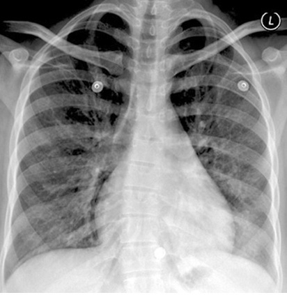
| PA View | Normal |
| Lateral Chest | Normal |
| Perfusion scan | L |
Other Cases of PE
| CXR | L |
| CXR | L |
| CT angiogram | L |
| CT angiogram | L |
| CT angiogram | L |
| Nuc Med | L |
| Angiography | L |
Pulmonary Embolism Images
CXR
Mostly normal or non specific
Triad: Basal infiltrate, Blunted costophrenic angle, elevated diaphragm
Wedge shaped density
Ventilation/Perfusion scan: Perfusion defects with normal vetilation
CT angiogram: Filling defects, Cut off of vessels
Pulmonary angiogram: Filling defects, Cut off of vessels