Genitourinary Imaging
Renal/Bladder Ultrasound
David A. Hatch, M.D.
Loyola University Stritch School of
Medicine
Ultrasound is performed by generating high frequency sound waves
(typically 5 - 10 kHz) and directing them through body tissues using
a probe held on the skin. The probe also contains a receiver to
detect sound waves (called echoes) reflected from tissues. When the
sound waves travel easily through uniform substances (water, oil,
urine, etc.), no echoes are generated. The ultrasound image seen on
the screen is, therefore, black; there are no echoes. When the sound
waves encounter a tissue that absorbs or transmits the sound, a wave
is reflected back to the probe. The ultrasound image is white or gray
depending on the intensity of the reflection. Unlike x-rays or CAT
scans, ultrasound doesn't detect tissue density. Rather, it detects
sonotransmission (the passage or reflection of sound). Highly dense
tissues such as bone or kidney stones readily reflect echoes and,
therefore, appear bright white on an ultrasound. Air, such as in the
bowel, also readily reflects echoes. The edge of the bowel,
therefore, appears white on an ultrasound. Therefore substances with
widely differing densities (air - bone) may both appear bright white
on an ultrasound. Remember, ultrasound does not detect tissue
density.
|
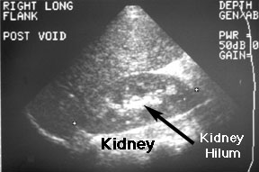
You need to know a few conventions used in ultrasound
imaging in order to read ultrasounds. First, try to get as
much information about the ultrasound image as you can from
the frame. You will see that this image has a label: "RIGHT
LONG FLANK POST VOID." This means that you are looking
at a longitudinal image of the right flank. The left
side of a longitudinal image is always the cephalad side of
the image. The right side of a longitudinal image is
the caudad side. The kidney is the bean shaped structure
marked at either end by a small cursor [+]. Superior to the
kidney and superficial (toward the top of the image) is the
liver. It is rather homogeneous (fairly regular grey
pattern). Notice that the kidney is not so homogeneous. In
the center of the kidney is an area of increased echoes
(light grey or white). This is the hilum of the kidney
also called the central sinus. It shows increased
echogenicity because there are several structures (pelvis of
the kidney, blood vessels, nerves, fat and lymphatics) that
transmit sound differently. As the sound wave hits the
interface between two such structures, an echo is generated.
|
|
|
This is a transverse image of a kidney. In a
transverse image, the left side of the image represents the
right side of the body, just as in a standard x-ray or
CT scan.
|
|
Usefulness: Ultrasound is the most useful pediatric
urologic imaging test. It is easily performed without any preparation
of the child and it causes no pain. It can differentiate solid from
cystic masses and it is very helpful in defining renal and bladder
anatomy. Using sophisticated computer analysis, current ultrasound
machines can detect movement. By this means they are able to detect
the velocity of blood flow in vessels. This is very useful in
allowing one to measure perfusion to a kidney or testis. Ultrasound
is also a relatively economical exam.
Limitations: Ultrasound detects shape and sonodensity. It
does not, however, measure function of kidneys. Nuclear renograms are
the best G/U imaging study to measure renal function.
Indications: Children with urine infections, abdominal
masses, suspected hydronephrosis or hematuria.
Examples: Normal kidney.
Hydronephrosis.
Kidney tumor.
Return to G/U Development Home
Page.
©David A. Hatch, M.D., 1996