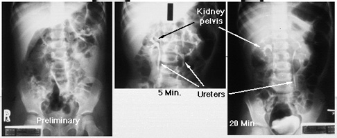
The preliminary film (on the left) is also called a 'scout film.' It is taken before intravenous contrast is given. This first image shows normal bones and soft tissues. Most kidney stones would be visible on the scout film. After contrast is injected intravenously, concentration of the contrast is seen in both kidneys (middle image - nephrogram phase). Contrast may also be seen in the proximal ureters. At 20 minutes after contrast injection (right image), urine containing contrast material is seen to drain through normal sized ureters into the bladder.
Return to G/U Imaging Home Page.
Return to G/U Development Home Page.
©David A. Hatch, M.D., 1996