You order the ultrasound seen on the right. Notice that the right kidney has hydronephrosis. The ureter is also dilated down to the pelvis and adjacent to the bladder. The ultrasonographer cannot find the point at which the ureter drains into the bladder. Why is this girl wet? Why does she have an abnormal right kidney?
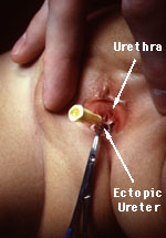
You examine the perineum carefully and find a small opening. You are able to pass a small catheter into the orifice and clear yellow liquid drains. After injecting contrast material into the catheter you obtain the x-ray seen on the right.
This girl has an ectopic ureter. Rather than draining into the bladder, this ectopic ureter drains directly onto the septum between the urethra and the vagina. Because it does not pass through the urethral sphincter, this ureter constantly drains. That is why this girl has never been dry.
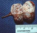
You tell to the mother that the child has an ectopic ureter. A renal scan confirms that the right kidney has minimal function. You surgically remove the dysplastic kidney seen on the right.
After you remove the kidney and ectopic ureter, Kelsey is completely dry. Her mother is delighted and very grateful. She recommends you to her close personal friends, Bill and Hillary Clinton, for a White House Fellowship.