|
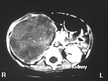
This is a contrast CAT scan; intravenous contrast was administered while the images were being obtained. Notice that the left kidney is relatively lighter that the adjacent muscle (posterior to the vertebral body) because contrast is being concentrated in the renal parenchyma. CAT scans can tell us if a kidney is perfused and if it has tubular function. If the kidney were not perfused and functioning, the renal parenchyma would be the same shade of gray as the adjacent muscle. Notice that the right kidney is enlarged. It doesn't have the uniform light gray that the left kidney has. Instead a relatively heterogeneous mass is present. Notice that there is a thin rim of white around the right kidney. This is normal renal parenchyma (concentrating the intravenous contrast) stretched over the encroaching tumor. This child has a Wilm's tumor, the most common renal tumor in children.
|
|