Ureterocele Case History 2
An 18-month-old Latino baby was brought to the emergency room
with fever to 104 degrees. Blood and urine cultures grew E. Coli.
After treatment with intravenous antibiotics imaging studies
were obtained.
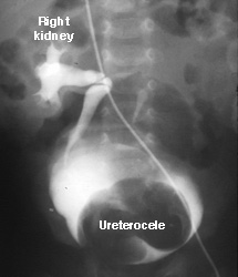
| This intravenous pyelogram (IVP) shows only one functioning kidney. The right renal pelvis and ureter are normal for a baby. Notice that the bladder is not uniformly filled with contrast. A large filling defect obscures the base and right side of the bladder. This is the dilated distal end of a left ureter, a ureterocele.
The left kidney actually functioned relatively well after the urine infection was completely treated. This study illustrates why an intravenous pyelogram is not a very sensitive indicator of renal perfusion and function. A renal scan would have shown perfusion and some limited function in the left kidney.
|  |
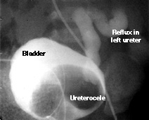
| A voiding cystourethrogram (shown at left) shows a filling defect (darker gray area) at the left bladder base. This is the ureterocele seen on the IVP. Notice that there is reflux into the left ureter. There are actually two separate ureters on the left side (complete duplication). We can tell this because there is no contrast in the ureterocele, but contrast does appear in a ureter. The contrast fills the lower pole ureter. The ureterocele is the distal end of the upper pole ureter. This ureterocele actually opened into the baby's urethra; it was an ectopic ureter. What does this mean about the function of the renal parenchyma associated with this upper pole ureter?
|  |
The ureterocele was opened and allowed to drain directly into
the bladder. A renal scan, however, later showed almost no function
in the upper pole of the kidney. Why?
Return to G/U Development home page.
©David A. Hatch, M.D., 1996