Intravenous Pyelogram of Horseshoe Kidney
This is an intravenous pyelogram (IVP) of a 10-year-old boy with a horseshoe kidney. The x-ray was obtained because he had hematuria after suffering a blow to the abdomen. Notice that the kidneys lie slightly lower in the abdomen than normal kidneys. As the fused lower poles of the horseshoe kidney ascend to the level of the inferior mesenteric artery, further ascent is halted.
| 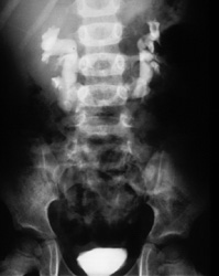 |
| Horseshoe kidneys have an abnormal axis. Draw a line between the uppermost calyx and the lowermost calyx on each side. In normal kidneys this line will be closer to the vertebral column at the upper pole. In horseshoe kidneys, the line will be closer to the vertebral column at the lower pole.
|  |
Horseshoe kidneys have a higher incidence of hydronephrosis and
renal stones than normal kidneys. Horseshoe kidneys usually have
multiple renal arteries. For
Return to Normal Renal Development.
Return to G/U Development home page.
©David A. Hatch, M.D., 1996