![]()
![]()
IMAGING:
Planar
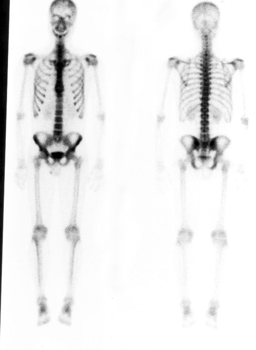
All whole body imaging
begins with planar images. Usually this is done with a camera capable of
obtaining an image of the whole body with one acquisition. Cameras with
two heads can obtain both an anterior and a posterior image simultaneously.
|
This is a normal whole body bone scan in a 27 y/o male patient. The anterior image is on the left and the posterior image is on the right. |
|
SPECT
SPECT (Single Photon Emission Computed Tomography) is a technique which allows imaging of organs or portions of the body in cross sectional planes. Normally the transaxial, coronal and sagittal planes are displayed, although the image can be reconstructed in a plane in any angle chosen by the operater.
|
These are examples of normal SPECT images of the lumbar spine in a 27 y/o male referred because of back pain. |
|
|
|
|
![]()
| Gary L Dillehay, M.D. |
Last Updated: August 15, 1996 |