Workup
Hematuria and Flank pain
What is the common cause for hematuria or flank pain?
What are the useful imaging modalities to investigate renal stones? Indicate when you would select each procedure. Utility of imaging procedures.
Controversy exists over the proper procedure sequence. With the addition of non-contrast helical CTs, some advocate CT as the first-line procedure. However, most physicians continue to utilize plain radiographs of the abdomen (KUB) as the initial screening imaging procedure.
- Abdominal radiograph of the kidneys, ureter, and bladder (KUB)
- Allows for the evaluation of urinary tract stones
- Ninety percent or more of urinary-tract stones are radio-opaque.
- The degree of opacity varies depending upon the composition of stone.
(calcium content of the stone)
- Uric acid stones (8%) are radio lucent.
- Visibility of stones depends not only on the degree of opacity, but also on their sizes and positions relative to other abdominal structures.
- An opaque stone needs to be approximately 2mm in its largest diameter to be visible on an abdominal film.
- Poor sensitivity for ureteral stones
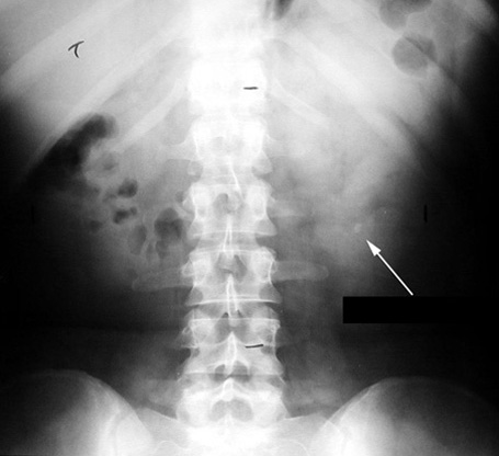 |
Kidney Stone
|
- Non-contrast helical computed tomography of the abdomen (NCHCT)
- Greater sensitivity than KUB for ureteral stones
- Most sensitive and specific (95-97% and 96-97% respectively)
- Because abdomen and pelvis are scanned in one to two breaths, virtually eliminates the problem of respiratory motion
- Can be performed quickly, eliminating potential delays
- Safe, because eliminates the risk of contrast, which includes allergic reactions and toxicity
- Detects non-urologic pathology – including appendicitis or ovarian cysts
- While more expensive than a KUB, comparable to a excretory urography and ultrasound
- Able to detect all stones regardless of composition, except those associated with indinavir therapy for HIV infections
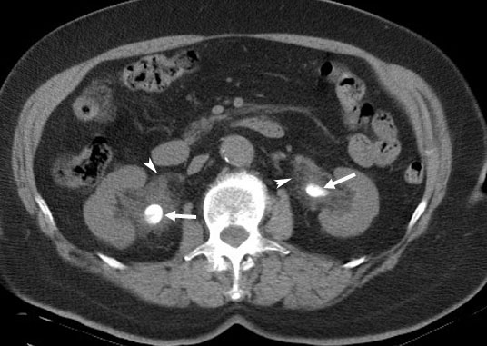 |
Bilateral Renal Pelvis Calculi
Arrowheads: There is soft tissue infiltration secondary to extravasation of urine around both renal pelvis. |
- Ultrasound
- Prefered procedure in preganant women and in patients allergic to IV
contrast.
- Not dependent on the composition of stones and detects uric-acid stones as well as calcium stones.
- Stones are seen as highly echogenic foci and often produce distal acoustic shadowing
- Not always possible to distinguish small stones from arterial calcifications, pericalceal fat, or crystal-laden calceal submucosal plaques.
- Detects hydronephrosis
- Generally good sensitivity .
- Ureteral stones are difficult because of overlying gas
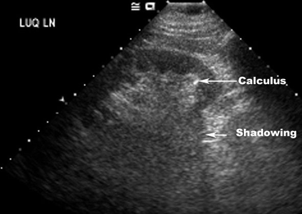 |
Kidney Stone
US: Lower pole calculus. |
Sensitivity, specificity of procedures.
PROCEDURE |
SENSITIVITY |
SPECIFICITY |
KUB |
45-70% |
77% |
CT |
95-97% |
96% |
Ultrasound |
32-70% |
70-97% |