Pleural effusion
Objectives:
Definition:
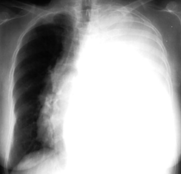
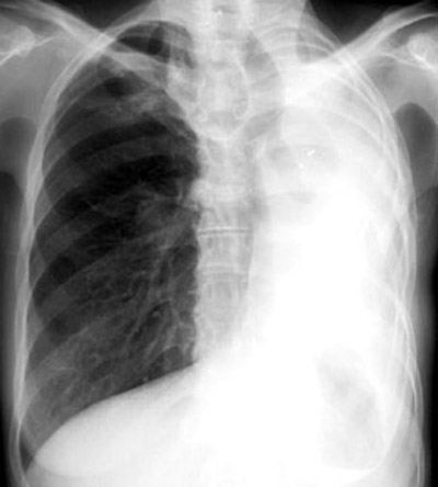
You are looking at pleural effusion on left, in the Chest x-ray.
Irrespective of the nature of fluid, radiologically they will look
similar.
Q1: What are the radiological criteria for pleural effusion?
Radiological criteria are:
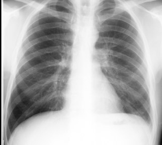
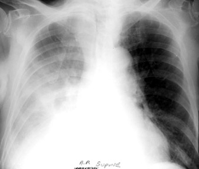
Q2: How do you differentiate complete opacification of one hemi thorax?
Consider
Mediastinum is shifted to opposite side with effusion and
pulled to same side with atelectasis
Hemithorax is larger with effusion and smaller with atelectasis
There are other reasons for loss of unilatweral lung volume, but for now
remembre atelectasis and resection.
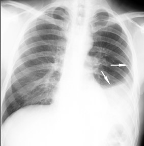
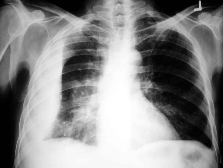
Q3: What are the different appearances of pleural effusion?
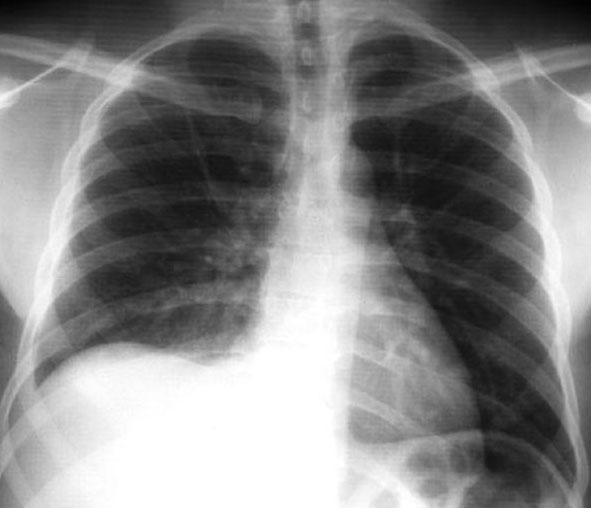
Q4: How do you recognize loculated effusion? What is the significance?
Q5: When will you order lateral decubitus film?
Lateral decubitus film is obtained
Most of the time it is ordered unnecessarily with no additional benefit in large effusions.
Q6: How do you determine the etiology of effusion from chest x-ray?
Radiologically you cannot distinguish transudate, exudate, blood or pus. It mainly the associated findings suggest the etiology. Let me give few scenarios
Q7: How does radiological procedures help in thoracentesis?
You do not need any radiological assistance to tap most of the effusions. Obtain radiological assistance to tap
Ultrasound is the preferred method for localiztion of fluid and a needle can be passed through the probe. It can also be done at the bed side if necessary. CT guided tap is equally effective, except it can not be useful for bed side tap.