| This exercise is for students studying Anatomy. Try answering the questions from the images before looking at the labeled images for answers. | |||
|
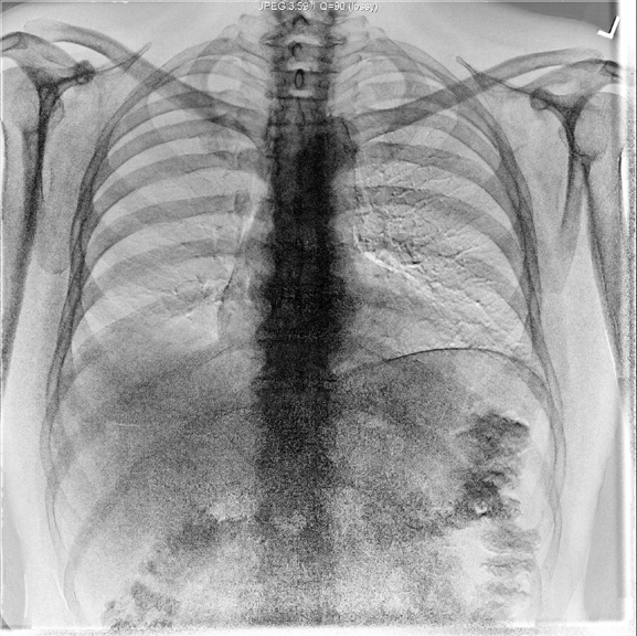
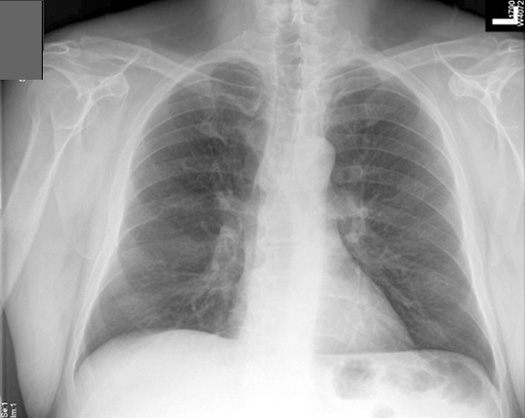
PA view |
|||
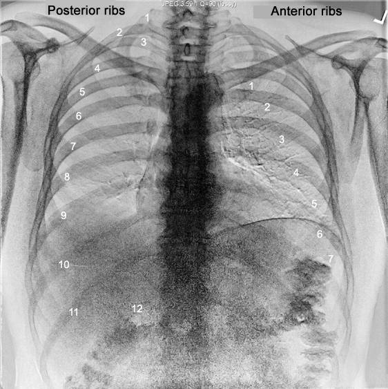
| CXR 1 | Identify the anterior and posterior ribs | Answer | |
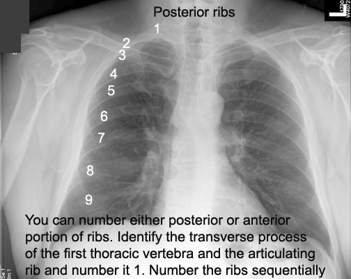
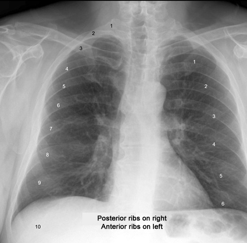
| CXR 2 | How do you number the posterior ribs? | Answer | |
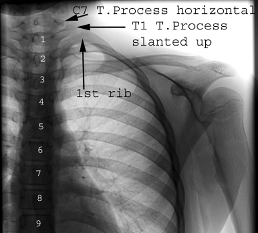
| CXR3 | How do you identify the first thoracic vertebra? | Answer | |
| CXR 4 | Number the anterior ribs? | Answer Answer | |
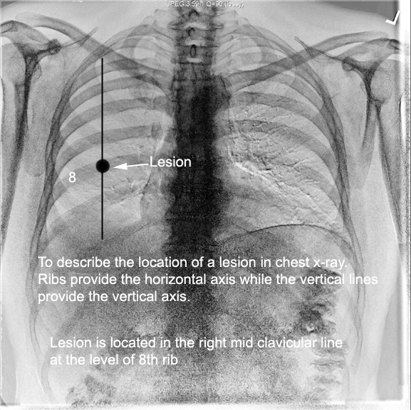
| What is the purpose of numbering ribs? | Answer | ||
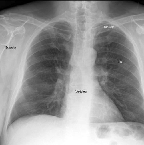
| CXR 5 | Identify Vertebra, scapula, ribs, clavicle | Answer | |
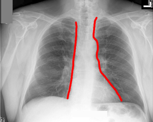
| CXR 6 | Draw the outline of Mediastinum. | Answer | |
| CXR 7 | What are the Images at which you measure mediastinal width? | Answer | |
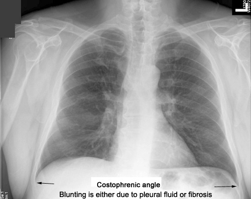
| CXR 8 | Identify costo diaphragmatic (costo phrenic) angles | What does blunting of costo diaphragmatic angle imply? | Answer |
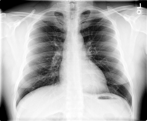
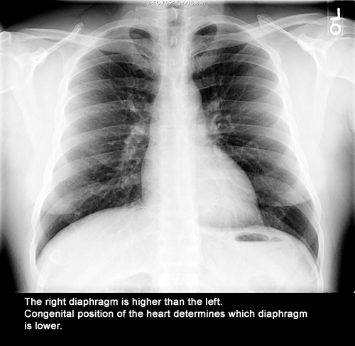
| CXR 9 | Identify diaphragm positions | Which diaphragm is higher and why? | Answer |
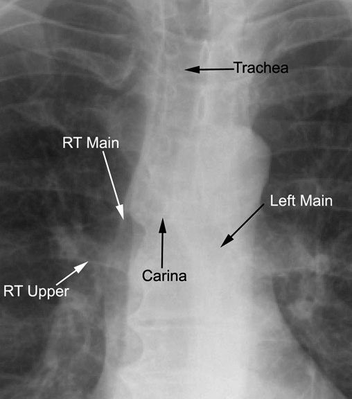
| CXR 10 | Identify Trachea, carina, right and left main stem bronchi. | Why are they visible, while rest of the bronchial tree is not? | Answer |
| CXR 11 | Identify right transverse fissure | What is the normal location? | Answer |
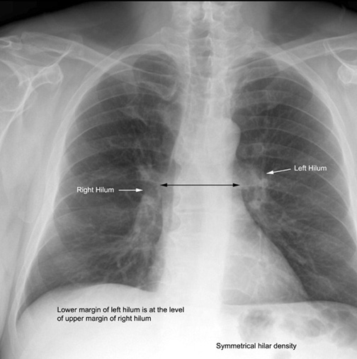
| CXR 12 | Identify left and right hilum. | What is the normal relationship? | Answer |
|
Lobar Projections |
|||
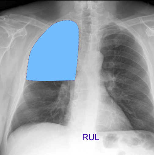
| CXR 1 | Visualize RUL projection in PA view | Answer | |
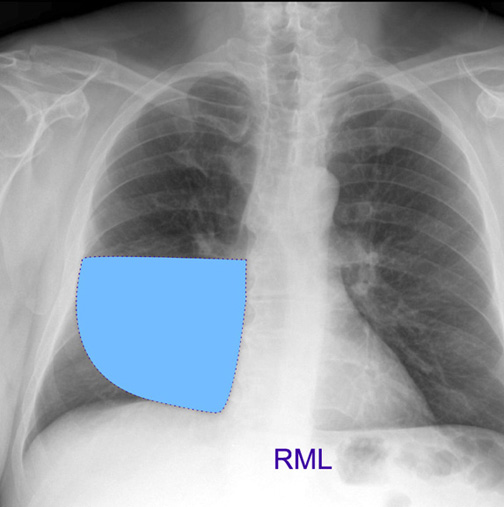
| CXR 2 | Visualize RML projection in PA view | Answer | |
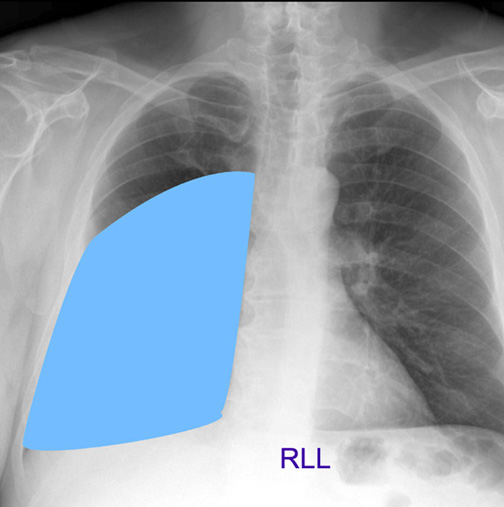
| CXR 3 | Visualize RLL projection in PA view | Answer | |
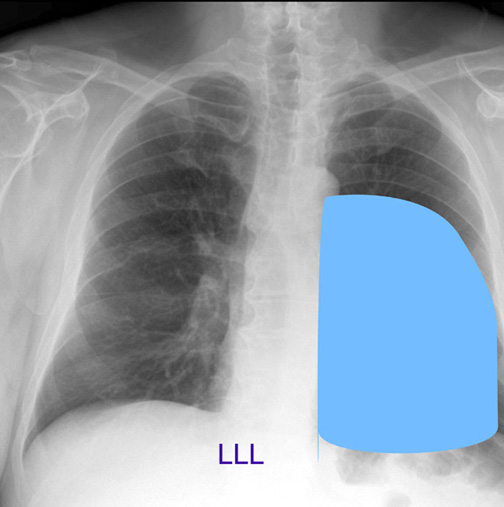
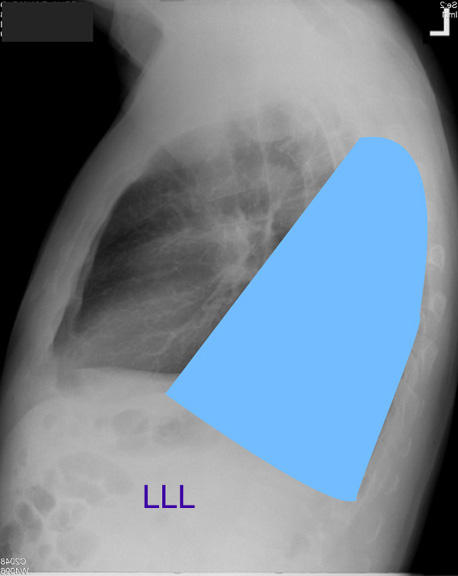
| CXR 4 | Visualize LLL projection in PA view | Answer | |
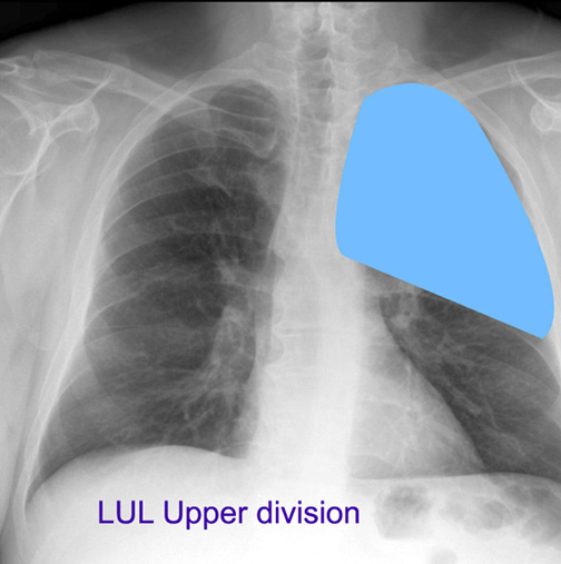
| CXR 5 | Visualize LUL projection in PA view | Answer | |
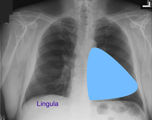
| CXR 6 | Visualize Lingula projection in PA view | Answer | |
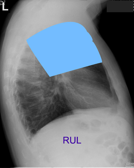
| CXR 7 | Visualize RUL projection in lateral view | Answer | |
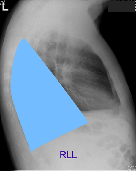
| CXR 8 | Visualize RLL projection in lateral view | Answer | |
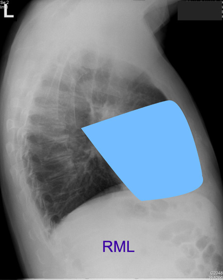
| CXR 9 | Visualize RML projection in lateral view | Answer | |
| CXR 10 | Visualize LLL projection in lateral view | Answer | |
|
Heart |
|||
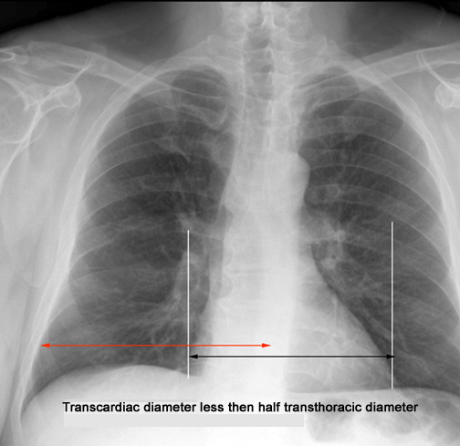
| CXR 1 | How do you measure heart size? | What is the normal range for heart size? | Answer |
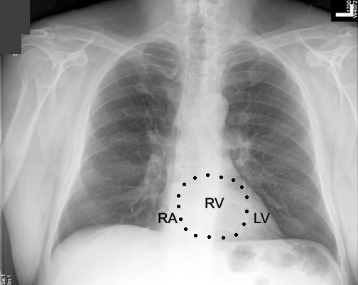
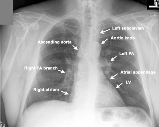
| CXR 2 | Identify chambers of heart in PA view | Answer | |
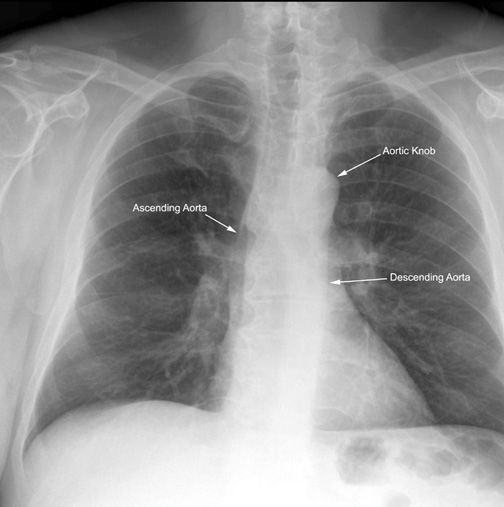
| CXR 3 | Identify ascending aorta, aortic knob and descending aorta | Answer | |
| CXR 4 | Identify structures along the edge of mediastinum | Answer | |
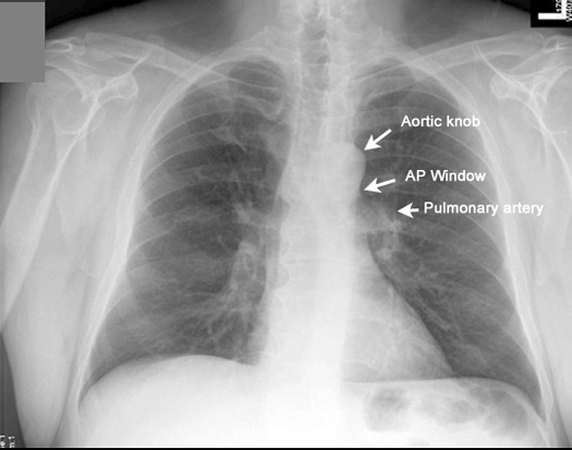
| CXR 5 | Identify Aortic knob, AP window and Pulmonary artery | What can fill AP window? | Answer |
|
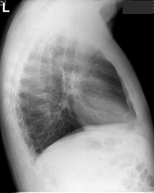
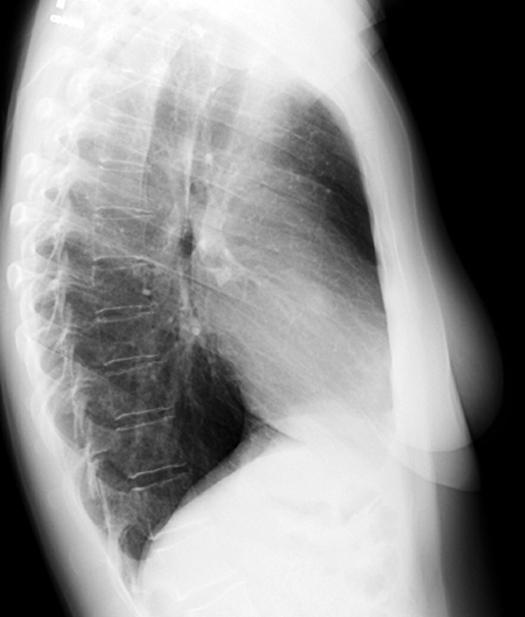
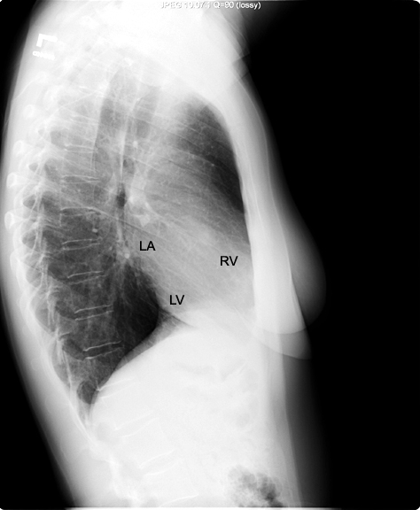
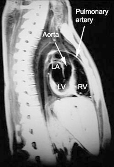
Lateral view |
|||
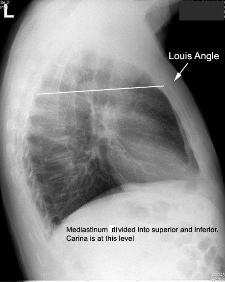
| CXR 1 | Identify Sternum and angle. of Louis. | What are the important landmarks at Sternal angle (Angle of Louis)? | Answer |
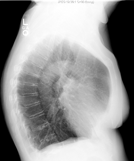
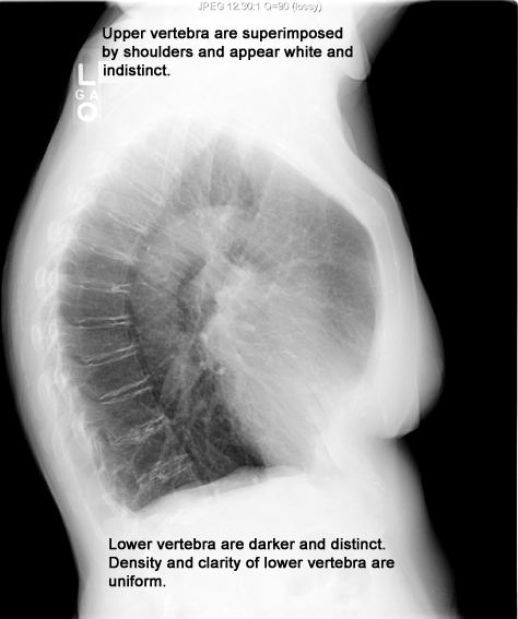
| CXR 2 | Identify Vertebra. | Answer | |
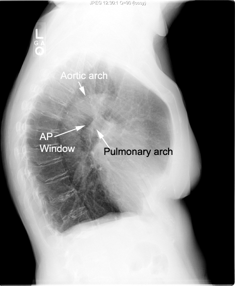
| CXR 3 | Identify aortic, pulmonary arches and AP window | Answer | |
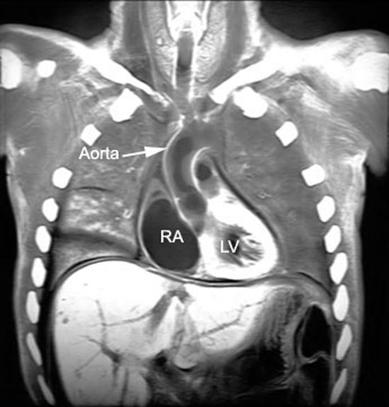
| CXR 4 | Identify chambers of heart in lateral view | Answer | |
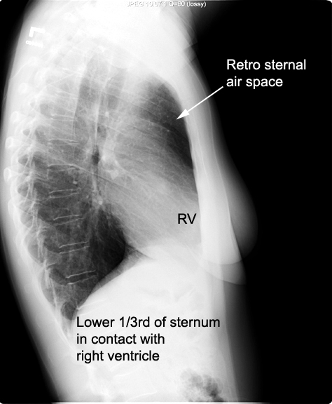
| CXR 5 | Identify retro sternal air space. | What is normal retro sternal space? | Answer |
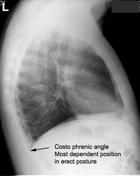
| CXR 6 | Identify costo diaphragmatic angle | How do we use this information? | Answer |
|
CT |
|||
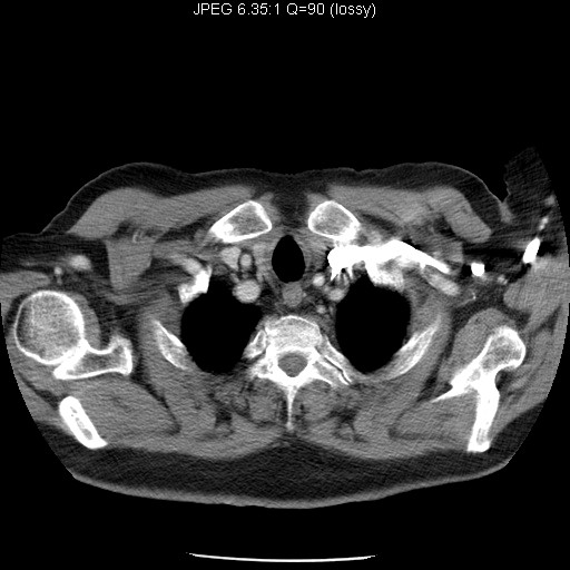
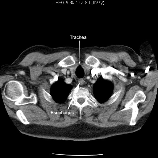
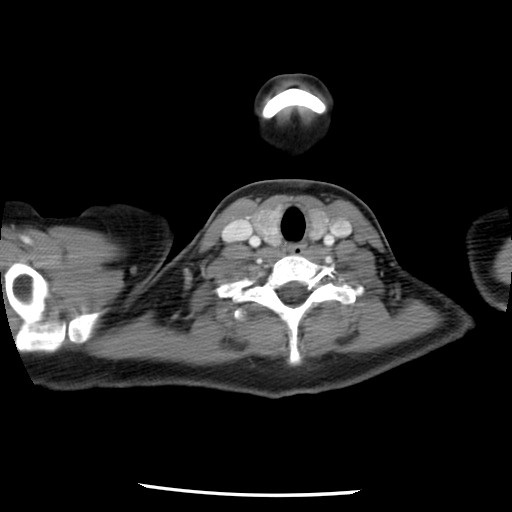
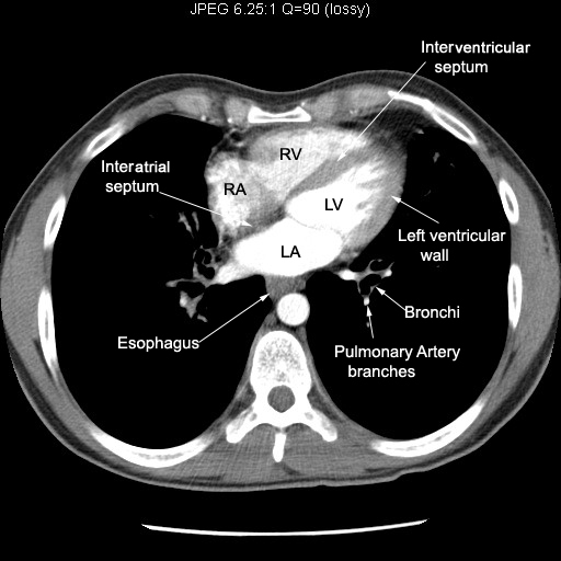
| Image 1 | Identify Trachea and Esophagus | Answer | |
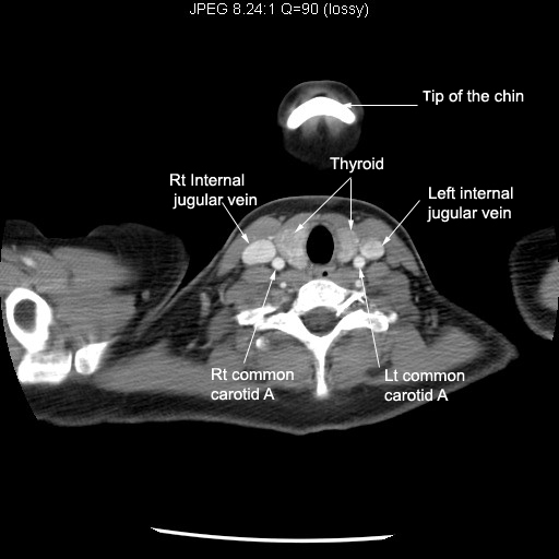
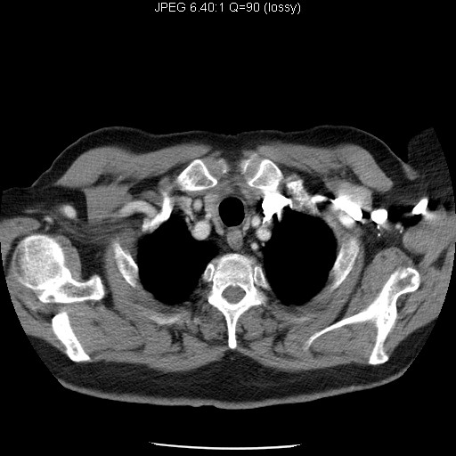
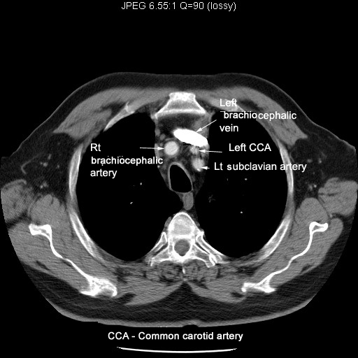
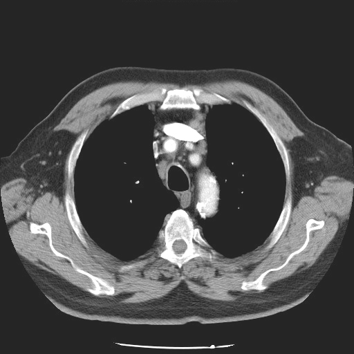
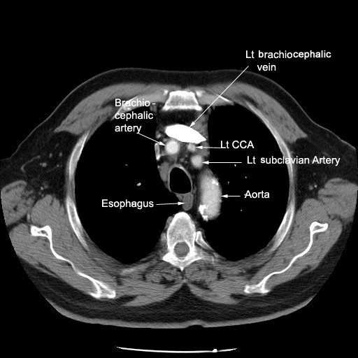
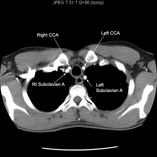
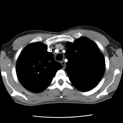
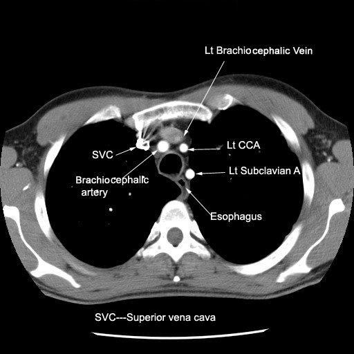
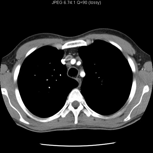
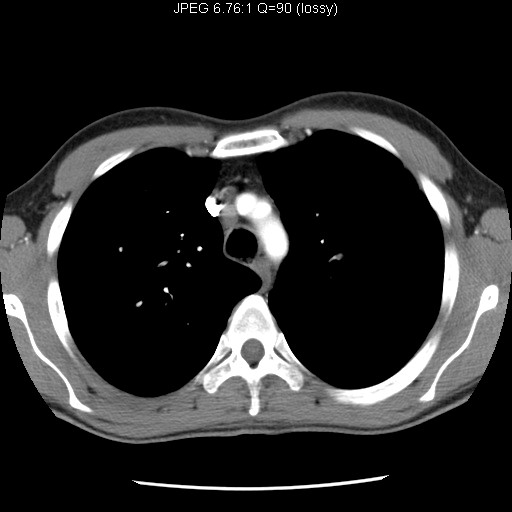
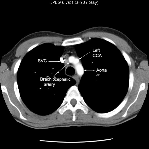
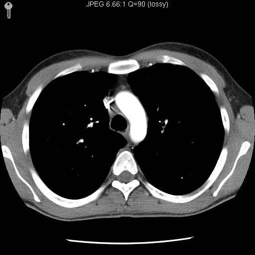
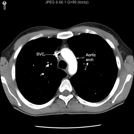
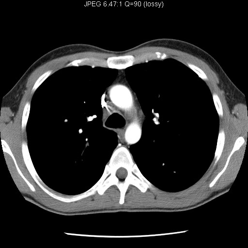
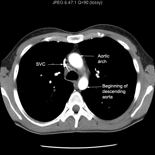
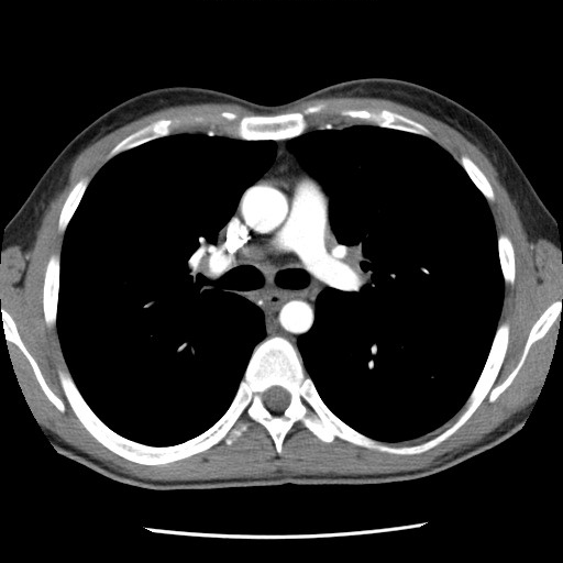
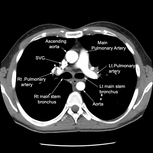
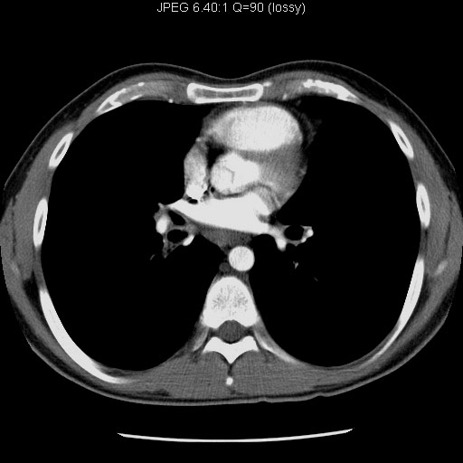
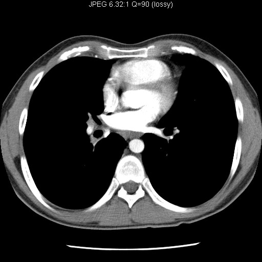
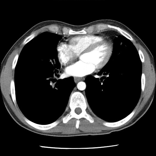
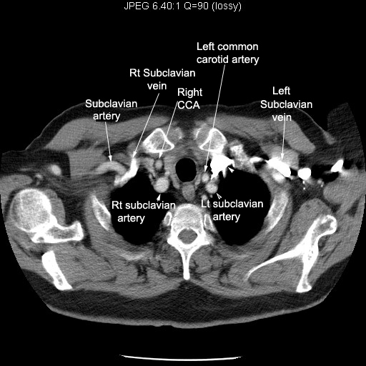
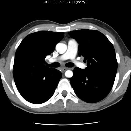
| Image 2 | Identify vascular structures | Answer | |
| Image 3 | Identify vascular structures | Answer | |
| Image 4 | Identify vascular structures | Answer | |
| Image 5 | Identify vascular structures | Answer | |
| Image 6 | Identify vascular structures | Answer | |
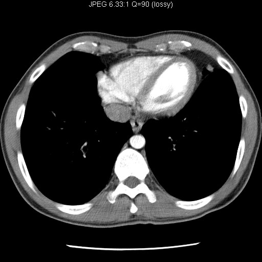
| Image 7 | Identify vascular structures | Answer | |
| Image 8 | Identify vascular structures | Answer | |
| Image 9 | Identify vascular structures | Answer | |
| Image 10 | Identify vascular structures | Answer | |
| Image 11 | Identify vascular structures | Answer | |
| Image 12 | Identify vascular structures | Answer | |
| Image 13 | Identify vascular structures | Answer | |
| Image 14 | Identify vascular structures | Answer | |
| Image 15 | Identify vascular structures | Answer | |
| Image 16 | Identify vascular structures | Answer | |
|
CT |
|||
| Major veins
|
Sequence1
Sequence2 Sequence3
Sequence4 Sequence5
Sequence6 Sequence7
Follow the major veins draining into superior vena cava. |
||
| SVC | Right and left brachiocephalic veins join to form superior vena cava. Follow the superior vena cava entering right atrium | ||
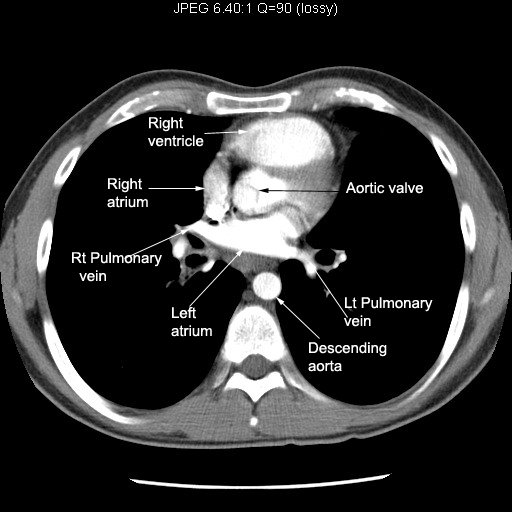
| Right atrium | Sequence
1 Sequence 2 Sequence
3 Sequence 4
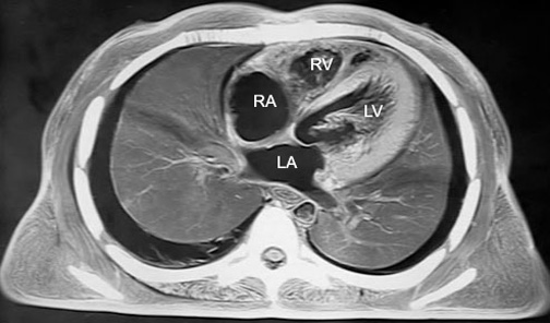
Follow SVC entering right atrium. Note the relationship of right atrium to other chambers of heart. |
||
| Right ventricle | Sequence 1 Sequence 2 Sequence 3 Sequence 4 Note the relationship of right ventricle to Sternum. |
||
| Pulmonary artery |
Follow pulmonary artery. Identify right and left branches. |
||
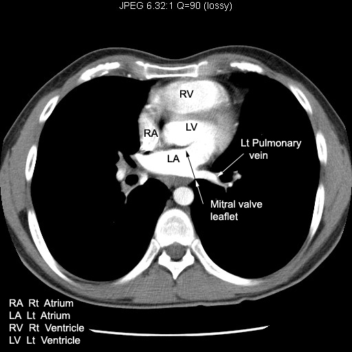
| Pulmonary veins | Sequence 1 Sequence 2 Sequence 3 Note Pulmonary veins entering left atrium. |
||
| Left atrium | Sequence 1 Sequence 2 Sequence 3 Note mitral valve leaflet. Note its location.. |
||
| Left ventricle |
Note left ventricular wall thickness and Inter ventricular septum |
||
| Aorta | Sequence 1 Sequence 2 Sequence 3 Sequence 4 Sequence 5 Sequence 6 Sequence 7 Sequence 8 Follow Aorta from head end to foot. Note major branches, ascending, arch and descending portions of Aorta. |
||
| Major branches | Sequence
1 Sequence 2 Sequence
3 Sequence 4
Follow the major branches of Aortic arch to neck. |
||