Plain x-ray
Principle
Examples
- Chest x-ray
- Abdomen
- Bones and Joints
How it is done
Example Indications
- Chest x-ray
- Most commonly used imaging
- Practically done routinely in most
of the hospitalized patients
- For evaluation of pulmonary or
cardiac symptoms
- Abdomen
- Bones and Joints
Limitation
- Significant pathology can be missed
Useful for
- It could be diagnostic
- It serves as preliminary image to plan for
more specific imaging studies
- Required for interpretation of other imaging
procedures Eg V/Q Lung scan
Cost: $
HIDA scan
- A HIDA scan is an imaging test used to examine
the gallbladder and the ducts leading into and out of the gallbladder.
- How it is done
- The patient receives an intravenous
injection of a radioactive material called hydroxy iminodiacetic acid (HIDA).
- The HIDA material is taken up by the liver
and excreted into the biliary tract.
- In a healthy person, HIDA will pass
through the bile ducts and into the cystic duct to enter the
gallbladder.
- It will also pass into the common bile
duct and enter the small intestine, from which it eventually makes its
way out of the body in the stool.
- HIDA imaging is done by a nuclear scanner,
which takes pictures of the patient's biliary tract over the course of
about two hours.
- The images are then examined by a
radiologist, who interprets the results.
- Safety
- It is generally a very safe test and is
well tolerated by most patients.
- Indication
- Usually, HIDA scans are ordered for
patients who are suspected of having an obstruction in the biliary
tract, most commonly those who are thought to have a stone blocking the
cystic duct leading out of the gallbladder.
- Such a scenario is consistent with acute
cholecystitis, which often requires surgical removal of the gallbladder.
- In cholecystitis, HIDA will appear in the
bile ducts, but it will not enter the cystic duct or the gallbladder --
a finding that indicates obstruction.
- If the HIDA enters the bile ducts but does
not enter the small intestine, then an obstruction of the bile duct
(usually due to stones or cancer) is suspected.
- Cost
Nuclear Medicine studies
Principle
How it is done
- Most of the studies are performed by injecting
a radioisotope into a vein.
- The type of isotope used varies with each
study
- The isotope flows through the blood vessels
and concentrates in the organ that is being tested
- Scanning of body or organ follows
- When to start and end scanning varies with
each study
Useful for
Example Indications
-
Renal scan: To evaluate Renal
function
-
Bone scan: To evaluate bone
metastasis
-
Perfusion lung scan: Suspected
patients with pulmonary embolism
-
Myoview: Suspected aptients with
coronary artery disease
-
Testicular scan: To evaluate
Testicular torsion
Advantages
- Radionuclide imaging is safe since it does not
carry the risk of allergic reaction encountered with contrast
- Radiation exposure is minimal.
Limitation
Radionuclide study
Principle
Examples
- Lung scan
- Renal scan
- Bone scan
How it is done
- A nuclear renogram is performed by
injecting a radioisotope into a vein.
- Scanning begins with injection and is
carried out up to 30 minutes.
- Delayed views may be necessary.
- The isotope flows through the blood
vessels of the kidney and is filtered by the glomerulus and secreted
by the renal tubules.
- As the isotope flows into the kidney, it
is detected by a nuclear medicine gamma camera usually placed
posterior to the kidneys.
- The amount of isotope filtered and
drained by the kidneys is analyzed by a computer.
- Perfusion, secretion and excretion of
the kidney are determined with this test.
Useful for
Example Indications
Advantages
- Radionuclide imaging is considered safe
since it does not carry the risk of allergic reaction encountered with
contrast
- Radiation exposure is minimal.
Limitation
Nuclear medicine Testicular scan
- This study is usually ordered on an
emergency basis
- 20 mCi of 99mTcO4 pertechnetate is given
I.V. (adult dose).
-
Scan for approximately 30 minutes.
- Useful to differentiate acute testicular
torsion from other causes of testicular pain/swelling.
- This study demonstrates the presence or
absence of perfusion to the testes.
- Flow is decreased or absent with
torsion
- Epididymitis and orchitis have
normal or increased perfusion.
Bone scan
- A bone scan is a test that detects areas of
increased or decreased bone metabolism (turnover).
- How it is done
- A radiotracer (bone-seeking radionuclide)
is injected into the bloodstream through a peripheral vein.
- As it decays, the radiotracer emits gamma
radiation, which is detected by a camera that slowly scans body.
- The camera is used to capture images to be
used to determine how much of the radiotracer collects in the bones.
- If a bone scan is performed to evaluate
possible fracture or infection, images will be performed shortly after
the radiotracer injection, as well as after a 3-hour delay, when the
tracer has collected in the bones.
- This is called a 3-phase bone scan.
- To evaluate metastatic bone disease,
images are obtained only after the 3-hour delay. Information from the
camera is recorded in a computer, which then processes the data and
creates an image.
- The scanning part of the test will last
about an hour and may require moving to various positions.
- Indication
- The test is performed to identify abnormal
processes involving the bone such as tumor, infection, fracture, or an
underlying metabolic disorder.
- Normal
- Normal distribution areas appear uniform
and gray throughout all the bones in body.
- What abnormal results mean
- There should be no areas of asymmetric
increased or decreased distribution of the radionuclide.
- "Hot spots" are areas where
there is increased bone uptake (accumulation) of the radiotracer; these
appear black.
- "Cold spots" are areas where
there is less uptake of the radiotracer.
- These appear light or white.
- Safety
- Cost
V/Q scan; Ventilation/perfusion
scan; Lung ventilation/perfusion scan
 A
pulmonary ventilation/perfusion scan is a pair of nuclear scan tests that
use inhaled and injected radioactive material (radioisotopes) to measure
breathing (ventilation) and circulation (perfusion) in all areas of the
lungs.
A
pulmonary ventilation/perfusion scan is a pair of nuclear scan tests that
use inhaled and injected radioactive material (radioisotopes) to measure
breathing (ventilation) and circulation (perfusion) in all areas of the
lungs.- How it is done
- A pulmonary ventilation/perfusion scan is
actually two tests that may be performed separately or together.
- The perfusion scan is performed by
injecting radioactive albumin into a vein.
- The patient is immediately placed on a
movable table that is positioned under the arm of a scanner.
- The lungs are scanned to detect the
location of the radioactive particles as blood flows through the lungs.
- The ventilation scan is performed by
scanning the lungs while having the person inhale radioactive gas.
- A mask is placed over the nose and mouth,
and the patient is asked to breathe the gas while sitting or lying on
the table beneath the arm of the scanner.
- A chest X-ray is usually performed prior
to or following a ventilation and perfusion scan.
- Indication
- The ventilation scan is used to evaluate
the ability of air to reach all portions of the lungs.
- The perfusion scan measures the supply of
blood through the lungs.
- A ventilation and perfusion scan is most
often performed to detect a pulmonary embolus.
- It is also used to evaluate lung function
in people with advanced pulmonary disease such as COPD and to detect the
presence of shunts (abnormal circulation) in the pulmonary blood
vessels.
- Normal
- A ventilation and perfusion scan should be
correlated with a chest X-ray.
- There should be uniform uptake of
radioisotope in all portions of the lungs with equal distribution in
both lungs.
- What abnormal results mean
- A decreased uptake of radioisotope during
a perfusion scan indicates a problem with blood flow, including
occlusion of the pulmonary arteries.
- A localized decreased in perfusion scan
uptake (particularly when ventilation scan is normal) may indicate
pulmonary embolus.
- Larger areas of decreased perfusion scan
uptake may indicate a condition such as pneumonitis.
- A decreased uptake of radioisotope during
a ventilation scan may indicate reduced breathing and ventilation
ability or airway obstruction.
- A decreased ventilation uptake (plus X-ray
evidence of consolidation) may indicate pneumonia.
- Larger areas of poor uptake may indicate
damage from chronic smoking or COPD.
- Safety
- Cost
CAT scan; Computed axial
tomography (CAT) scan
Principle
- CT scanning combines X-rays and computer to
produce precisely detailed cross-sectional images of the organs.
- A thin x-ray beam rotates around the patient.
- Detectors measure the amount of x-rays
that make it through particular area of interest.
- CAT scans can be enhanced by using intravenous
iodinated contrast material.
- Normal enhancement of parenchyma can be
compared to increased enhancement of tumor..
- When necessary, three-dimensional models of
organs can be created
Examples
- Chest CT
- Head CT
- Abdomen CT
- Joint CT
- Bone CT
How it is done
- The patient lies on a
narrow table that slides into the center of the scanner.
- IV access will be required if contrast
media needs to be administered
- Subject will be required to hold breath and not
move as motion causes blurred images.
- Scan time varies depending on the type of
scanner and required studies.
- The newest multidetector scanners can
image from head to toe, in less than 30 seconds.
- Spiral scanners can
perform the examination in one continuous motion of the gantry.
Useful for
- CT scan is helpful in delineating the precise anatomy
of organs
- Three-dimensional reconstructions studies and
blood supply provide "road maps" for planning surgeries.
Example Indications
- CT is often utilized in the trauma setting
to evaluate the brain, chest, and abdomen.
- CT chest is done routinely in patients
with Lung cancer to evaluate the mass and nodes
- CT can be used to guide
interventional procedures, such as biopsies and placement of drainage
tubes
Limitation / Safety
- Table cannot fit obese patients
- Some patients cannot lie supine or stay still
- Contrast allergy issues
Cost: $$
Magnetic resonance imaging;
Nuclear magnetic resonance (NMR) imaging
- A powerful magnet generates a magnetic field
roughly 10,000 times stronger than the natural background magnetism from the
earth.
- A very small percentage of hydrogen atoms
within a human body will align with this field.
- When focused radio wave pulses are broadcast
towards the aligned hydrogen atoms in tissues of interest, they will return
a signal.
- The subtle differences in that signal from
various body tissues enables MRI to differentiate organs, and potentially
contrast benign and malignant tissue.
- Any imaging plane (or "slice") can
be projected, stored in a computer, or printed on film. MRI can easily be
performed through clothing and bones.
-
- How it is done
- An MRA, or magnetic resonance angiogram, is a
special type of MR that creates three-dimensional reconstructions of vessels
containing flowing blood and is often utilized when conventional angiography
cannot be performed due to renal failure or other contraindications
- Indication
- Normal
- Abnormal
- Safety
- Cost
MRI (Magnetic
Resonance Imaging)
Principle
- MRI uses a strong magnet, radio waves
and computers to create detailed images of the body.
- It does not use ionizing radiation
unlike conventional radiography and Computed
Tomographic (CT) imaging
- MRI imaging is based on the magnetic properties of atoms.
Examples
- Head MR
- Abdomen MR
- Joint MR
- Bone MR
How it is done
- Patient lays inside a massive hollow magnet,
and is exposed to short bursts of powerful non-ionizing radio
wave energy, directed at protons, the nuclei of hydrogen or water
atoms, in the body.
- Radio signals generated by first
"exciting" and then "relaxing" those protons, are
computer-processed to form digital images, reflecting different types
of tissue.
- Scanner
must be located within a specially shielded room to avoid outside
interference.
- The patient will be asked to lie on a
narrow table which slides into a large tunnel-like tube within the
scanner.
- Special body coils will be placed
around the areas to be
studied.
- These special body coils send
and receive the radio wave pulses, and improve the
quality of the images.
Advantages
Example Indications
Limitation
- Expensive
- Limited availability
- A complete scan, depending on the sequences performed,
may take
up to one hour or more.
- Newer scanners may complete the
process in less time.
- Certain types of metals (Heart valves) can cause significant errors in the reconstructed images.
- MRI has limited applicability for the
urinary tract since the non-specificity of its signals makes it
ineffective in detecting calcifications and bladder abnormalities.
Ultrasound (Sonogram)
Principle
- The use of high-frequency sound
waves to produce real-time images, provides a simple and painless way to
examine structures.
- An ultrasound machine sends out high-frequency sound
waves, which reflect off body structures.
- A computer receives these reflected waves
and uses them to create a picture.
How it is done
- A clear, water-based conducting gel is
applied to the skin over the area being examined to help with the
transmission of the sound waves.
- A hand held Transducer is
then moved over the area being examined.
- Transducer sends high
frequency sound waves into the body.
- The waves are reflected back by various
tissues they go through.
- The reflected waves , with a help of a
computer, form an image on the screen.
- Color coding of the various reflected
echoes gives color images.
Doppler
- Doppler examination is done using US
waves aimed at a moving object - arteries or veins.
- The reflected
waves with computer aid, give us the velocity of blood in various
vessels.
Example Indications
- Pregnancy evaluation
- Ectopic pregnancy
- Tortion Scrotum
Advantages
- Non-invasive test
- Requires no preparation
- No pain
- Provides accurate anatomic information,
including dimensions
- No radiation risk
- Avoiding the potential allergic and
toxic complications of contrast media.
- Can be used on individuals with poor
kidney function in whom contrast cannot be given
- No complications
- Can be done at bedside
- Relatively economical exam
Cost: $
Limitation
Duplex/Doppler ultrasound
- These
tests measure blood flow and blood pressure.
- A duplex study is a test which uses Doppler
ultrasound to assess and/or monitor blood flow through arteries or
veins.
- Plethysmography measures changes in blood
volume in a blood vessel.
- How it is done
- For the Duplex/Doppler ultrasound:
- Clothing from the area being monitored
is removed, and a probe with a conductive gel (like vasoline) on the
tip is placed on various points along the vessel being tested.
- This enables the technician to
evaluate blood flow through the vessels.
- The information is relayed to the
ultrasound monitor to be viewed and recorded.
- For a plethysmography:
- Blood pressure is first monitored in
both arms.
- The clothing from the extremity being
tested is removed and the patient lies on his or her back.
- A blood pressure cuff is applied to
the extremity being tested and inflated until the pulsatile flow is
no longer heard.
- Then the pressure is released from the
cuff until the flow returns.
- The blood pressure when flow returns
is recorded and the information is transmitted to a computer which
records and interprets the information.
- The blood pressure cuff may be moved
to other positions on the same or other extremities during the
test.
- After the readings are taken, the
patient may be re-tested while in another position such as sitting
or standing.
- Indication
- These tests are noninvasive (external)
tests used to determine if there is significant disease in either
arteries or veins, if adequate blood is reaching an extremity, to
evaluate trauma to a blood vessel, or to monitor patients with arterial
reconstruction or graphs.
- These tests can also detect blood clots.
- Normal
- As part of a duplex ultrasound, the doctor may
calculate an ABI or ankle-brachial index.
- This number is obtained by dividing the
Doppler or systolic pressure of the ankle by the pressure in the arm.
- A value of 0.9 or greater is normal.
- Your doctor will also evaluate the flow of
blood in the vessels with the ultrasound.
- Safety
- Cost
Transthoracic echocardiogram (TTE);
Echocardiogram - transthoracic; Doppler ultrasound of the heart; Surface echo
- Echocardiogram is a test that uses sound waves
to create a moving picture of the heart.
- The picture is much more detailed than X-ray
image and involves no radiation exposure.
- How it is done
- A trained sonographer performs the test,
then your physician interprets the results.
- An instrument that transmits
high-frequency sound waves called a transducer is placed on your ribs
near the breast bone and directed toward the heart.
- The transducer picks up the echoes of the
sound waves and transmits them as electrical impulses.
- The echocardiography machine converts
these impulses into moving pictures of the heart.
- Echocardiogram works well for most
patients and allows doctors to see the heart beating and to visualize
many of the structures of the heart.
- Occasionally, because your lungs, ribs, or
body tissue may prevent the sound waves and echoes from providing a
clear picture of heart function, the sonographer may administer a small
amount of a dye through an IV to better see the inside of the heart.
- Very rarely, more invasive testing using
special echocardiography probes may be necessary.
- If the echocardiogram is unclear due to a
barrel chest, congestive obstructive pulmonary disease, or obesity, your
health care provider may choose to perform a transesophageal
echocardiogram, or TEE. With TEE, the back of your throat is
anesthetized and a scope is inserted down your throat.
- On the end of the scope is an ultrasonic
device that an experienced technician will guide down to the lower part
of the esophagus, where it is used to obtain a more clear
two-dimensional echocardiogram of your heart.
- Indication
- This test is performed to evaluate the
valves and chambers of the heart in a noninvasive manner.
- The echocardiogram allows doctors to
evaluate heart murmurs, check the pumping function of the heart, and
evaluate patients who have had heart attacks.
- It is a very good screening test for heart
disease in certain groups of patients.
- Normal
- A
normal echocardiogram reveals normal heart valves and chambers and
normal heart wall movement.
- Safety
- Cost
RNV; Cardiac blood pooling
imaging; Nuclear heart scan; Radionuclide ventriculography; MUGA
- A test that uses radioactive tracers to
delineate heart chambers and major blood vessels leading to and from the
heart.
- The procedure is non-invasive
- The heart structures are not touched by
instruments.
- How it is done
- A radioactive isotope is injected into the
vein.
- Commonly used isotopes include technetium
and thallium.
- Radioactive isotopes attach to red blood
cells and pass through the heart in the circulation.
- The radioactive isotope can be traced
through the heart using special cameras or scanners
- The images may be synchronized with an
electrocardiogram.
- The test is often given at rest then
repeated with exercise or after administering certain medications.
- The test may be performed to detect a
heart attack, to evaluate those at risk of coronary artery disease
without invasive testing (coronary angiography and heart
catheterization), and to evaluate heart wall motion and pumping function
of the heart.
- Normal
- Normal results indicate normal heart valve
and chamber structure and function, or a normal cardiac response to
exercise.
- Safety
- Cost
Cardiac angiography; Angiography -
heart
- Coronary
angiography is a procedure in which a contrast material that can be seen
using X-ray equipment is injected into one of the arteries of the
heart.
- This allows you to view the flow of blood
through your heart.
- How it is done
- Coronary angiography is usually performed
in conjunction with cardiac catheterization.
- Patient will be given a mild sedative
prior to the test to help you relax.
- The study is carried out in a laboratory
by a trained cardiologist or radiologist and technicians or
nurses.
- An intravenous (IV) line is inserted into
one of the blood vessels in your arm or groin after the site has been
cleansed and numbed with a local anesthetic.
- A catheter is then inserted through the IV
and into your blood vessel.
- The catheter is carefully threaded into
the heart using an X-ray machine that produces real-time pictures
(fluoroscopy).
- Once the catheter is in place, contrast
material is injected and pictures are taken.
- Coronary angiography is performed to
detect obstruction in the coronary arteries, which can lead to heart
attack.
- Indication
- It may be performed if you have unstable
angina, atypical chest pain, aortic stenosis, or unexplained heart
failure.
- The test may also be performed for other
reasons.
- Normal
- Adequate blood supply to the heart is a
normal finding with a coronary angiogram.
- Safety
- Cost
- A Cardiac Cathetherization (also called a
heart cath or heart angiogram) is a test to show the inside of your heart
and coronary blood vessels. A long, thin, tube is placed in a blood vessel
in your groin or arm and advanced toward the heart. Once it is in the right
location in the heart or blood vessel, a dye is injected and xrays and
videos are taken. The test is done by a cardiologist (heart specialist) who
is assisted by nurses and technicians in the Cardiac Cath lab. The test
takes about 1 hour.
Angiogram; Angiography
- Arteriography
is a procedure in which a contrast material that can be seen using X-ray
equipment is injected into one of the arteries
- How it is done
- An arteriogram can be used to examine
almost any artery, including those of the head, kidneys, heart, or
lungs.
- The study is carried out in a laboratory
by a trained cardiologist or radiologist and technicians or nurses.
- First the doctor will need insert an
intravenous (IV) line into one of the blood vessels in your arm, chest,
neck, or groin.
- A catheter is then inserted through the IV
and into your blood vessels using an X-ray machine that produces
"live" pictures.
- Once the catheter is placed into the blood
vessel of interest, contrast material is injected and pictures are
taken.
- An arteriogram can be used to examine almost
any artery, including those of the head, kidneys, heart, or lungs.
- Cerebral angiography (head)
- Extremity angiography (arm or leg)
- Renal angiography (kidneys)
- Pulmonary angiography (lungs)
- Lymphangiography (lymph vessels)
- Right heart ventriculography (looking at
the right side of the heart)
- Left heart ventriculography (looking at
the left side of the heart)
- Coronary angiography (looking at the
vessels of the heart)
- Aortic angiography or aortography (looking
at the aorta, the major artery from the heart)
- Eye angiography
- Cardiac catheterization
- Indication
- The reasons for this test depend on the
type of arteriogram that will be performed.
- In general, arteriograms give the best
pictures of the body's blood vessels.
- Arteriograms are used to make specific
diagnoses and to help determine what the best treatment is in a
particular case.
- Often, the treatment itself can be
performed using the same type of catheters used in the arteriogram,
instead of requiring a more extensive surgery in an additional
procedure.
- It is sometimes used as part of a
procedure to repair the blood vessels called balloon angioplasty.
- Safety
- Cost
Angiogram
CT and MRI have replaced the
need for renal angiogram. It is rarely done nowadays for diagnostic
purposes..
However the therapeutic
applications of angiography have expanded considerably.
Principle
Angiography provides a
complete examination of the arterial supply to organs, including a
view of the aorta and branches.
Examples
- Aortogram
- Cerebral angiogram
- Renal angiogram
- Pulmonary angiogram
How it is done
- A catheter is inserted into abdominal
Aorta and contrast dye is injected into desired arteries.
- X-ray images are taken at a rapid rate
because the high pressure of the arterial blood flow will cause the
dye to disappear quickly.
Useful for
Past indications
- For evaluation of renal artery stenosis
- For delineation of vascular renal tumors
- For evaluation of tumor invasion of
renal veins and inferior vena cava
Limitation
- Invasive procedure
- Bleeding and injury to the artery.
- Contrast complications
Contrast filling lumens
Principle
Examples
- GI studies
- Bronchogram
- Cystogram
- Retrograde pyelogram
- Fistulogram
How it is done
Useful for
Example Indications
Advantages
Limitation
Principle
How it is done
Useful for
Example Indications
Advantages
Limitation
Contrast in blood stream
Principle
Examples
How it is done
Useful for
Example Indications
Advantages
Limitation
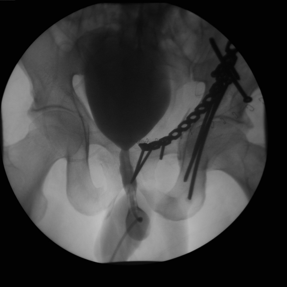
Voiding Cystourethrogram
A VCUG uses X-rays and
iodinized contrast to study the bladder and urethra.
- A voiding cystourethrogram
is performed by inserting a catheter into the urethra.
- Contrast material is then instilled into
the bladder through the catheter.
- X-rays are taken before, during and
after filling of the bladder.
- When the bladder is full, the catheter
is removed.
- While the patient
voids, additional x-rays are taken.
- Children with urinary tract infections.
- Reflux
is detected if contrast is seen to flow in retrograde fashion up
the ureters from the bladder.
- Pelvic trauma where rupture of the
bladder or urethra is suspected.
- If the bladder is ruptured, extravasation
of contrast will be seen outside the bladder in the pelvis or
abdomen.
- If urethra is ruptured, there is
extravasation into the perineum.
- Patients with suspected bladder outlet
obstruction
- Obstructions or strictures or injury
of the urethra can be seen on the x-rays taken during voiding.
Limitations:
- Insertion of the catheter is painful.
- While conventional voiding cystograms
are still necessary to evaluate the male urethra for posterior valves
and bladder trauma, the majority of reflux studies today are done
effectively with radionuclide cystography.
Retrograde Urethrogram
- Preliminary film of the urethra and
bladder is obtained prior to injection of a contrast agent
- A catheter is inserted about two
centimeters into the penis.
- The balloon on the catheter is slightly
inflated and contrast is injected.
- Several X-rays are obtained during the
injection of the contrast.
- The bladder is then filled and the
catheter is usually removed.
This is commonly performed in
patients in whom trauma to the penile urethra is suspected.
Limitation
- Minor discomfort during and following
the procedure
- Risk of contrast allergy
- Risk of urinary tract infection
Retrograde Pyelogram
- Defines ureters and collecting systems
- While newer diagnostic techniques have
replaced this test for many functions, retrograde pyelography may
still yield better definition of the upper urinary tract, particularly
the ureter and kidney collecting system.
How is retrograde pyelogram done?
- Urologist performs cystoscopy first
- A catheter is inserted inot ureteral
orifice and contrast dye is injected.
- This technique produces definitive
images of calcifications and tumors thus allowing easy diagnosis of
obstructions in the urinary system.
- Commonly performed when IVP produces an
inadequate picture. Useful to study urinary tract obstruction when
further clarification of nature of ureteral obstruction is required
- It also complements cystoscopy while
investigating a patient with hematuria or recurrent or suspected
cancer.
- Detects small lesions in the collecting
system E.g. Transitional cell carcinoma
Limitations
- Contrast complications
- May aggravate an existing urinary tract
infection or triggering one from the catheterization.
Scrotal Ultrasound
Scrotal ultrasound is used to
evaluate almost all abnormalities in the scrotum.
IVP (Intravenous pyelogram)
Principle
-
IVP is a radiological test
that uses contrast to outline the kidneys, ureters and bladder.
-
Also known as intravenous
urogram (IVU)
How it is done
Useful for
- Useful for evaluating the anatomy of the
kidneys, ureters and bladder
- One can detect function when no contrast
is excreted
- absence of renal function .
- absence of perfusion to a kidney
- Useful to identify urinary tract
obstruction
- Useful to evaluate reno-vascular
disease
Disadvantages
- Labor and time intensive – it may take
up to 6 hours to complete in the severe obstruction
- It requires placement of an intravenous
line.
- Requires a bowel preparation for optimal
results
- Involves intravenous injection of
potentially allergic and mildly nephrotoxic contrast
- Nonionic contrast agents have
lowered the incidence of adverse reactions.
- IVP's are not useful in patients with
renal dysfunction.
- Newborns rarely have sufficient renal
concentrating ability to allow the kidneys to be seen on an x-ray.
- Hydration is important
- May aggravate renal failure
Color Doppler US Scrotum
Color-Doppler imaging has made
ultrasonography a highly accurate and specific test for testicular torsion
What Is a DEXA Scan?
A Bone Density or DEXA scan uses x-rays to measure the amount of minerals
in your bones.
What Is a Bone Scan? A bone scan
shows any damage or activity within your bones by using a small amount of
radioactive material which is taken up by the bones. A scanner produces a
picture that can show very small amounts of changes in the bone. The radioactive
material is eliminated from your body very rapidly.
What is a F-18 FDG study?
A F-18 FDG study shows how different organs and
tissues in the body us (metabolize) sugar (glucose).
What Is a Lung Scan? A lung scan shows the
pattern of blood flow to lungs by using a small amount of radioactive material
injected into vein.
What Is a MUGA Study? A MUGA scan
shows how your heart is working. By using a small amount of radioactive
material, the camera can produce a picture that shows the heart at work.
What Is a Myoview Exercise Stress Test?
The Myoview Stress Test shows how much blood is being supplied to your heart
muscle while at rest and during exercise.
What Is a Adenoscan-Myoview Stress Test?
The Adenoscan - Myoview Stress Test shows how much blood is being supplied to
your heart muscle at rest and later while you receive a medication (Adenoscan)
through an intravenous line.. The test is for people who are unable to perform
an exercise stress test on a treadmill. This medication increases your heart's
blood flow as if you were exercising.
What is a Prostascint Scan? A Prostascint
scan can detect cancer cells in/and around the prostate gland or cells that
spread from the prostate. A special radioactive substance is injected into a
vein and is taken up by prostate cancer cells. The cells then can be detected
96-120 hours after the injection.
What is a Renal Scan with Lasix? A
Renal Scan with Lasix helps to decide how well kidneys are working and if
they are obstructed.
What is a Renal Scan with Vasotec? This
Nuclear Medicine test helps decide if altered blood flow to kidney is the cause
of your high blood pressure.
What is a Renal Scan with Lasix? A
Renal Scan with Lasix helps to decide how well kidneys are working.
What Is an Angiogram?
An angiogram is a test that evaluates blood vessels. To outline the blood
vessels so they can be seen on x-ray, a solution containing iodine (called
contrast media) is injected into a blood vessel. As the blood and contrast flows
through the smaller blood vessels, x-rays are taken.
What Is a Barium Enema?
A Barium Enema is a study of the colon or large bowel. A solution of barium is
given as an enema to coat the bowel walls and x-ray images or pictures are
taken.
What Is an Air Contrast Barium Enema?
An Air Contrast Barium Enema is a study of the colon or large bowel. A solution
of barium is given as an enema to coat the bowel walls; then air is injected to
distend the colon, and x-ray images or pictures are taken.
What Is a Chest X-Ray?
A Chest x-ray produces a picture of the lungs,
heart, windpipe and ribs.
What is a CT Scan? A CT Scan is a series of
computerized x-rays taken by a CT scanner, which is a specialized x-ray machine.
The scanner resembles a large doughnut with an examination table through the
center. The patient lies on the table and when the scans are taken, the table
moves slowly through the scanner. Scans can be done on any part of the body.
Common CT scans are of the chest, abdomen, pelvis, head spine and or blood
vessels. These scans allow doctors to see inside some areas of the body, which
cannot be seen using conventional x-rays.
What is a Cystogram/Voiding Cystogram?
A Cystogram/Voiding Cystogram is an x-ray test which shows the urinary bladder
and urethra by using a contrast media (a liquid containing iodine which outlines
the organs). This contrast material will be inserted into the bladder by a
radiologist through a catheter. The catheter is usually inserted through the
urethra by the radiologist or nurse. Several images will be taken.
What is an IVP (Intravenous Pyelogram)?
An intravenous pyelogram (IVP) is an x-ray test which examines the kidneys,
ureters and bladder by using contrast media (a a liquid which contains iodine
and is injected into a blood vessel). Several pictures or images will be
taken.
What Is a Mammogram? A mammogram is an
x-ray of the breast. The exam is performed using a special machine in which the
breast can be positioned to obtain a clear picture.
What Is a Myelogram? A myelogram is
an x-ray examination of the spinal canal. A contrast solution, containing
iodine, is injected into the fluid around the spinal cord. This solution
outlines the spinal cord so an image of it can be taken.
What is an UGI? An UGI (Upper
gastrointestinal) is a study of the esophagus and the stomach (the upper part of
the digestive system). A solution of barium is swallowed to coat the organs so
that they can be seen, and x-ray images or pictures are taken to show the lining
of the stomach and esophagus.
What is an UGI? An UGI/Small Bowel (Upper
GastroIntestinal/Small Bowel) is a study of the esophagus and the stomach (the
upper part of the digestive system) and the small bowel. A solution of barium is
swallowed to coat the intestines so that they can be seen, and x-ray images or
pictures are taken to show the movement of the solution through the intestines.
What Is an Ultrasound? An
ultrasound is a test that uses sound waves to "see inside" body. The
ultrasound does NOT use any radiation, special dyes or medications. Almost any
organ of the body can be "seen" by ultrasound.