Radiological procedures
Plain x-ray
Principle
Examples
How it is done
Useful for
Example Indications
Limitation
CT (Computerized
Tomogram)
Principle
CT scanning combines X-rays and
computer to produce precisely detailed cross-sectional images of the
organs.
Examples
- Chest CT
- Head CT
- Abdomen CT
- Joint CT
- Bone CT
How it is done
- A computerized axial tomography scan is
performed by obtaining axial x-rays images using computerized
reconstruction.
- CAT scans can be enhanced by using
intravenous iodinated contrast material.
- This allows one to detect perfusion
and concentration of the contrast by the kidney.
- Evaluates increased enhancement of
tumor with contrast separating it from normal enhancement of
parenchyma.
Useful for
- CT scan is helpful in delineating
the characteristics of anatomy and function of organs
- Three-dimensional reconstructions
studies and blood supply provide "road maps" for planning
surgeries.
Example Indications
Limitation
Ultrasound
Principle
The use of high-frequency sound
waves to produce real-time images, provides a simple and painless way to
examine structures.
Examples
How it is done
- A hand held transducer sends high
frequency sound waves into the body.
- The waves are reflected back by various
tissues they go through.
- The reflected waves , with a help of a
computer, form an image on the screen.
- Color coding of the various reflected
echoes gives color images.
- Doppler examination is done using US
waves aimed at a moving object - arteries or veins. The reflected
waves with computer aid, give us the velocity of blood in various
vessels.
Useful for
Example Indications
Advantages
- Non-invasive test
- Requires no preparation
- No pain
- Provides accurate anatomic information,
including dimensions
- No radiation risk
- Avoiding the potential allergic and
toxic complications of contrast media.
- Can be used on individuals with poor
kidney function in whom contrast cannot be given
- No complications
- Can be done at bedside
- Relatively economical exam
Limitation
MRI (Magnetic
Resonance Imaging)
Principle
- MRI uses a strong magnet, radio waves
and computers to create detailed images of the body.
Examples
- Head MR
- Abdomen MR
- Joint MR
- Bone MR
How it is done
- Lying inside a massive hollow magnet, a
patient is exposed to short bursts of powerful non-ionizing radio
wave energy, directed at protons, the nuclei of hydrogen or water
atoms, in the body.
- Radio signals generated by first
"exciting" and then "relaxing" those protons, are
computer-processed to form digital images, reflecting different types
of tissue.
- It does not use ionizing radiation.
Useful for
MRI is as good as CT or better
in characterizing lesions of organs
Example Indications
Limitation
- Expensive
- Limited availability
- MRI has limited applicability for the
urinary tract since the non-specificity of its signals makes it
ineffective in detecting calcifications and bladder abnormalities.
Angiogram
CT and MRI have replaced the
need for renal angiogram. It is rarely done nowadays for diagnostic
purposes..
However the therapeutic
applications of angiography have expanded considerably.
Principle
Angiography provides a
complete examination of the arterial supply to organs, including a
view of the aorta and branches.
Examples
- Aortogram
- Cerebral angiogram
- Renal angiogram
- Pulmonary angiogram
How it is done
- A catheter is inserted into abdominal
Aorta and contrast dye is injected into desired arteries.
- X-ray images are taken at a rapid rate
because the high pressure of the arterial blood flow will cause the
dye to disappear quickly.
Useful for
Past indications
- For evaluation of renal artery stenosis
- For delineation of vascular renal tumors
- For evaluation of tumor invasion of
renal veins and inferior vena cava
Limitation
- Invasive procedure
- Bleeding and injury to the artery.
- Contrast complications
Radionuclide study
Principle
Examples
- Lung scan
- Renal scan
- Bone scan
How it is done
- A nuclear renogram is performed by
injecting a radioisotope into a vein.
- Scanning begins with injection and is
carried out up to 30 minutes.
- Delayed views may be necessary.
- The isotope flows through the blood
vessels of the kidney and is filtered by the glomerulus and secreted
by the renal tubules.
- As the isotope flows into the kidney, it
is detected by a nuclear medicine gamma camera usually placed
posterior to the kidneys.
- The amount of isotope filtered and
drained by the kidneys is analyzed by a computer.
- Perfusion, secretion and excretion of
the kidney are determined with this test.
Useful for
Example Indications
Advantages
- Radionuclide imaging is considered safe
since it does not carry the risk of allergic reaction encountered with
contrast
- Radiation exposure is minimal.
Limitation
Nuclear medicine Testicular scan
- This study is usually ordered on an
emergency basis
- 20 mCi of 99mTcO4 pertechnetate is given
I.V. (adult dose).
-
Scan for approximately 30 minutes.
- Useful to differentiate acute testicular
torsion from other causes of testicular pain/swelling.
- This study demonstrates the presence or
absence of perfusion to the testes.
- Flow is decreased or absent with
torsion
- Epididymitis and orchitis have
normal or increased perfusion.
Contrast filling lumens
Principle
Examples
- GI studies
- Bronchogram
- Cystogram
- Retrograde pyelogram
- Fistulogram
How it is done
Useful for
Example Indications
Advantages
Limitation
Principle
How it is done
Useful for
Example Indications
Advantages
Limitation
Contrast in blood stream
Principle
Examples
How it is done
Useful for
Example Indications
Advantages
Limitation
Voiding Cystourethrogram
A VCUG uses X-rays and
iodinized contrast to study the bladder and urethra.
- A voiding cystourethrogram
is performed by inserting a catheter into the urethra.
- Contrast material is then instilled into
the bladder through the catheter.
- X-rays are taken before, during and
after filling of the bladder.
- When the bladder is full, the catheter
is removed.
- While the patient
voids, additional x-rays are taken.
- Children with urinary tract infections.
- Reflux
is detected if contrast is seen to flow in retrograde fashion up
the ureters from the bladder.
- Pelvic trauma where rupture of the
bladder or urethra is suspected.
- If the bladder is ruptured, extravasation
of contrast will be seen outside the bladder in the pelvis or
abdomen.
- If urethra is ruptured, there is
extravasation into the perineum.
- Patients with suspected bladder outlet
obstruction
- Obstructions or strictures or injury
of the urethra can be seen on the x-rays taken during voiding.
Limitations:
- Insertion of the catheter is painful.
- While conventional voiding cystograms
are still necessary to evaluate the male urethra for posterior valves
and bladder trauma, the majority of reflux studies today are done
effectively with radionuclide cystography.
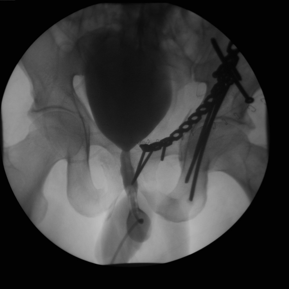
Retrograde Urethrogram
- Preliminary film of the urethra and
bladder is obtained prior to injection of a contrast agent
- A catheter is inserted about two
centimeters into the penis.
- The balloon on the catheter is slightly
inflated and contrast is injected.
- Several X-rays are obtained during the
injection of the contrast.
- The bladder is then filled and the
catheter is usually removed.
This is commonly performed in
patients in whom trauma to the penile urethra is suspected.
Limitation
- Minor discomfort during and following
the procedure
- Risk of contrast allergy
- Risk of urinary tract infection
Retrograde Pyelogram
- Defines ureters and collecting systems
- While newer diagnostic techniques have
replaced this test for many functions, retrograde pyelography may
still yield better definition of the upper urinary tract, particularly
the ureter and kidney collecting system.
How is retrograde pyelogram done?
- Urologist performs cystoscopy first
- A catheter is inserted inot ureteral
orifice and contrast dye is injected.
- This technique produces definitive
images of calcifications and tumors thus allowing easy diagnosis of
obstructions in the urinary system.
- Commonly performed when IVP produces an
inadequate picture. Useful to study urinary tract obstruction when
further clarification of nature of ureteral obstruction is required
- It also complements cystoscopy while
investigating a patient with hematuria or recurrent or suspected
cancer.
- Detects small lesions in the collecting
system E.g. Transitional cell carcinoma
Limitations
- Contrast complications
- May aggravate an existing urinary tract
infection or triggering one from the catheterization.
Scrotal Ultrasound
Scrotal ultrasound is used to
evaluate almost all abnormalities in the scrotum.
IVP (Intravenous
pyelogram)
Principle
-
IVP is a radiological test
that uses contrast to outline the kidneys, ureters and bladder.
-
Also known as intravenous
urogram (IVU)
How it is done
Useful for
- Useful for evaluating the anatomy of the
kidneys, ureters and bladder
- One can detect function when no contrast
is excreted
- absence of renal function .
- absence of perfusion to a kidney
- Useful to identify urinary tract
obstruction
- Useful to evaluate reno-vascular
disease
Disadvantages
- Labor and time intensive – it may take
up to 6 hours to complete in the severe obstruction
- It requires placement of an intravenous
line.
- Requires a bowel preparation for optimal
results
- Involves intravenous injection of
potentially allergic and mildly nephrotoxic contrast
- Nonionic contrast agents have
lowered the incidence of adverse reactions.
- IVP's are not useful in patients with
renal dysfunction.
- Newborns rarely have sufficient renal
concentrating ability to allow the kidneys to be seen on an x-ray.
- Hydration is important
- May aggravate renal failure
Color Doppler US Scrotum
Color-Doppler imaging has made
ultrasonography a highly accurate and specific test for testicular torsion