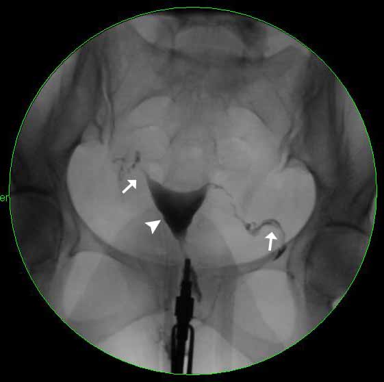
Image 1
This image shows an early radiograph from a normal HSG. The white arrowhead is pointing towards the uterus, which is filled with contrast (the radiopaque material), and is normal in shape. The white arrows are pointing towards the right and left fallopian tubes which are both beginning to fill with contrast.
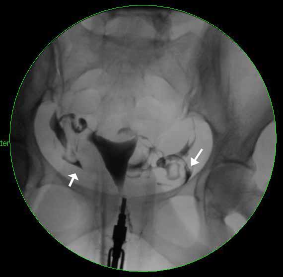
Image 2
The white arrows are pointing towards the dark, radio dense material that is spilling from both the right and left fallopian tubes. The arrows are pointing towards triangular shaped areas of contrast, which represents extravasation of into the peritoneum. It forms a triangular shape as it flows around bowel loops. This image represents a normal HSG.
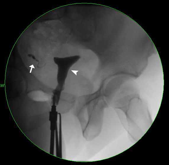
Image 3
The white arrowhead is pointing towards the uterus, which has filled with contrast and is normal in shape. The white arrow is pointing towards the right fallopian tube, which has begun to fill with contrast. Note that the left fallopian tube is not filling with contrast, which makes this an abnormal HSG.
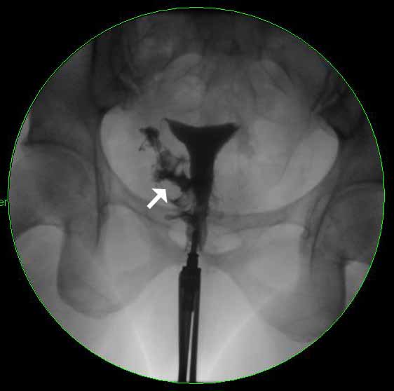
Image 4
At a later point in time, this image shows contrast spilling from the right fallopian tube into the peritoneum (white arrow). Note that the left fallopian tube did not fill with contrast and that there is no spillage on the left side which indicates that the tube is not patent.