|
|
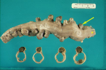
A section of colon reveals numerous diverticula which protrude from the edge of the taenia coli (*). The colon is cut in cross section revealing the diverticuli (contain feces) and the empty colonic lumen. |
|
|
A section of colon reveals numerous diverticula which protrude from the edge of the taenia coli (*). The colon is cut in cross section revealing the diverticuli (contain feces) and the empty colonic lumen. |
What is diverticulosis and what is diverticulitis?
What is the mode of clinical presentation of diverticulosis?
What are the potential complications of diverticula?
What are the complications of diverticulitis?
|
|
| Pathology | Imaging |
| Diverticula out pouching from colon | Diverticula in lower GI and CT scan |
| Diverticulitis | |
|
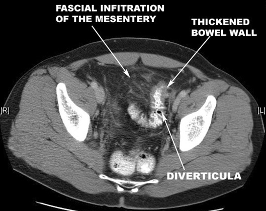
Facial infiltration of mesentery |
|
|
Ileus |
|
Thickened bowel wall |
| Complications | |
|
Inflammatory Mass with air pockets |
|
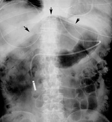
Air under diaphragm |
|
Fistulous tracks |
|
Nuclear medicine GI bleeding study |
|
Large bowel obstruction |
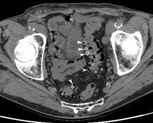 |
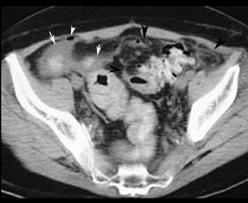
DiverticulaCT: Arrowheads point to multiple diverticula arising from the recto sigmoid. The contrast in diverticula is left over from previously administered GI contrast.
|
 |
|
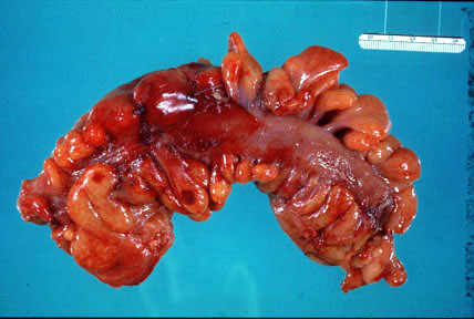
| Acute Diverticulitis: A section of colon reveals acute inflammation (hyperemia, swelling) of the serosa and pericolic fat. |
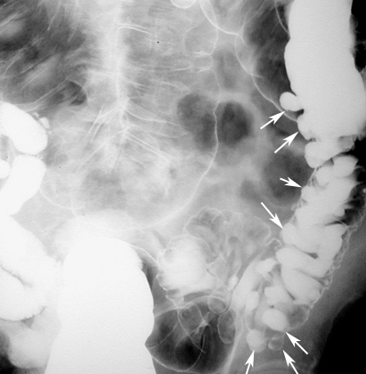
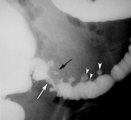
Lower GI showing Diverticula |
 |
 |
|
CT scan with GI contrast study showing findings of diverticulitis. |
|
 |
 |
Pneumoperitoneum
|
Pneumoperitoneum secondary to perforation of diverticulum.
|
|
|
|
|
|
|