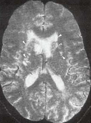
Cryptococcal Meningitis
Axial T2 Wtd image of the brain
Findings: Multiple small areas of increased T2 Wtd signal within both basal ganglia (arrows)
|
Cryptococcal MeningitisAxial T2 Wtd image of the brain Findings: Multiple small areas of increased T2 Wtd signal within both basal ganglia (arrows) |
Next |