Medullary Sponge Kidney
Pathology
- Congenital defect
- Manifest symptomatically in third or fourth
decade
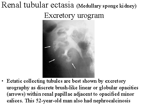
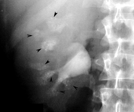
- Multiple cystic dilatation of the collecting
ducts in the medulla, diameters ranging from 1 to 5 mm.
- Not all papillae are equally involved
- Bilateral in 70% of cases
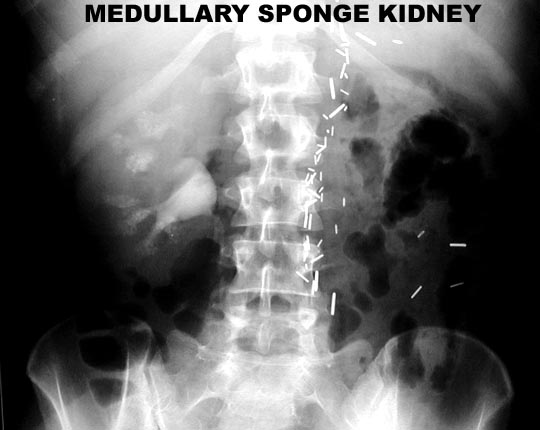
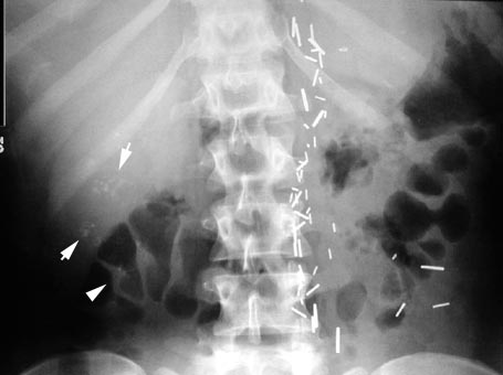
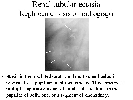
- Calculi are frequently found in the dilated
collecting ducts
Imaging strategy
- Patients present with renal stones or
infection or hematuria or discovered incidentally
- Most commonly diagnosed by intravenous
pyelogram.
- In the modern days IVP is not done any more.
- About 60% have renal
stones
- Plain film and CT
abdomen are the imaging procedures of choice for investigation of patients
suspected to have renal stones.
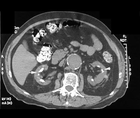
- In CT you may see bilateral renal
calcifications in the cortico medullary
junction
ADPKD
Renal
cancer MSK