Esophagus
- General
- Course
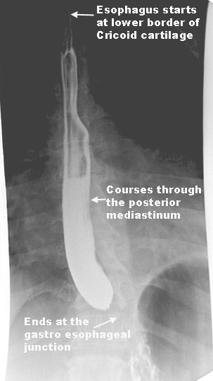
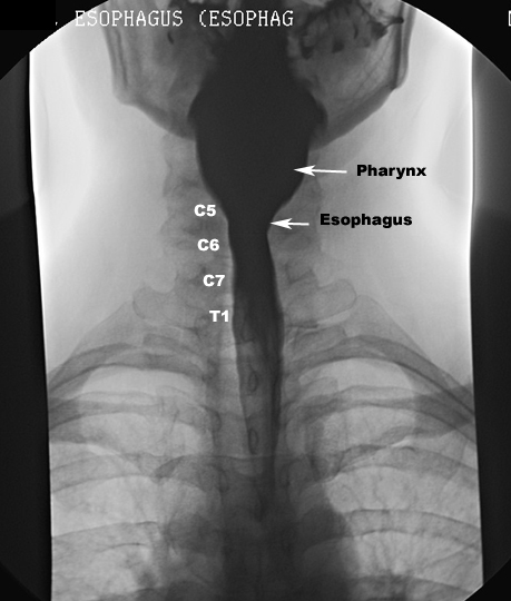
- It begins in the neck at the lower border
of the cricoid cartilage, opposite the sixth cervical vertebra, descends
in front of the vertebral column, through the superior and posterior
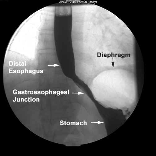
mediastinum, passes through the diaphragm, and entering the abdomen,
ends at the cardiac orifice of the stomach, opposite the eleventh
thoracic vertebra
- The general direction of the esophagus is
vertical, but it presents two slight curves in its course
- Just before it perforates diaphragm it
presents a distinct dilatation
- We need to consider its relationship to
structures at different levels
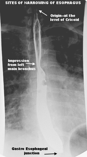
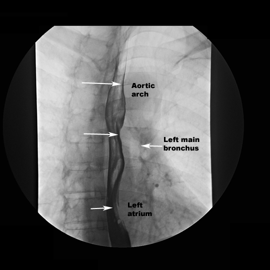
- On Left anterior oblique view of the Barium
swallow, following impressions are seen on the esophagus:
- Aortic arch
- Left main bronchus
- Left atrium
- Relations
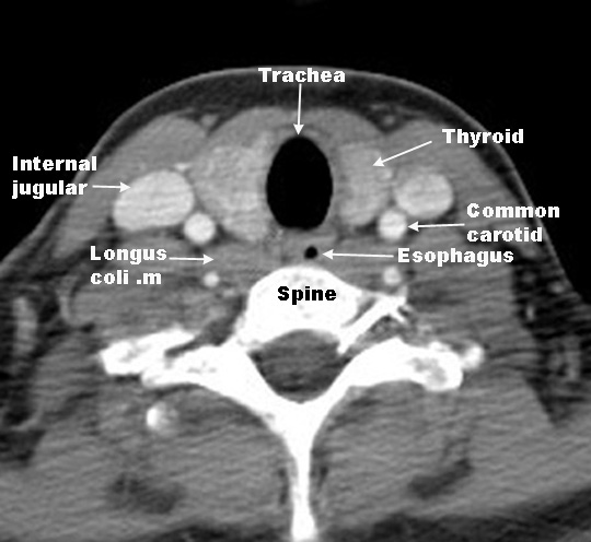
- The cervical portion
- Anterior
- Posterior
- Vertebral column
- Longus coli muscles
- Either side
- Common carotid artery
- Thyroid gland
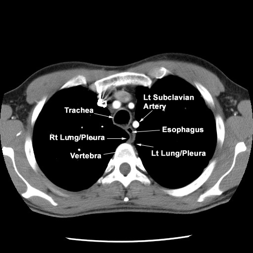
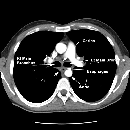
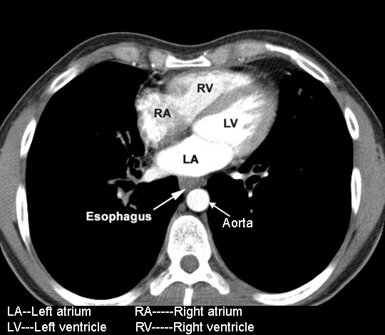
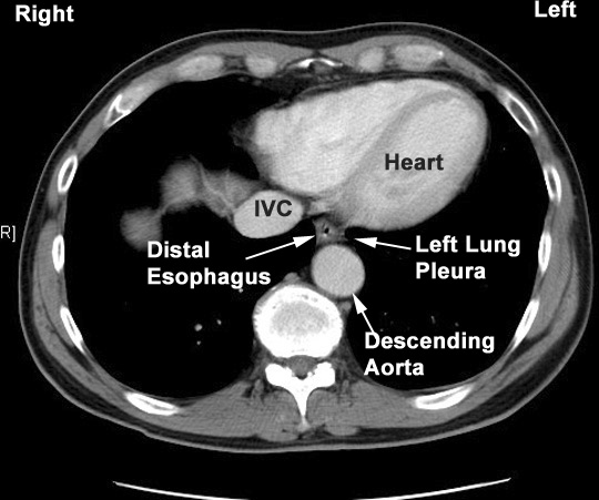
- The thoracic portion Superior
Mediastinum Superior
Mediastinum At Carina At
Heart Distal
- Anterior
- Trachea
- Left main stem bronchus
- Pericardium
- behind and to the right of aortic
arch
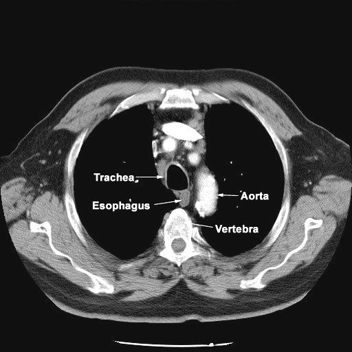
- Posterior
- Vertebral column
- Hemiazygous vein
- right aortic intercostal arteries
- aorta near diaphragm
- Right
- right pleura
- azygous vein
- Left
- aortic arch
- left subclavian artery
- left pleura
- descending aorta
- Abdominal portion
- Lies in the esophageal grove on the
posterior surface of the left lobe of the liver
- It measures about 1.25 cm in length
- It is somewhat conical with its base
applied to the upper orifice of the stomach, and is known as the
antrum cardacum
Applied anatomy
Why do you have to know the sites of narrowing in
Esophagus?
Which pleural space will be infected in
Esophageal tear?
Which cardiac chamber when enlarged can
indent and displace Esophagus?
How do you recognize dilatation of esophagus in
chest x-ray?