REACTION TO INJURY: REPAIR
The hepatocyte has a florid regenerative potential.In experimental partial resection in the rat,the remaining liver tissue starts to regenerate within a few hours.In 14-15 hours DNA replication is seen.In 20-21 hours mitoses appear.In 32 hours mitoses are at the pick.In 2 weeks the remaining liver tissue has reached the weight that had before resection. Cell division takes place in the periportal zone.In pathological conditions,dead liver cells are replaced by proliferation of surviving liver cells.Hepatocytes,Kupffer cells,endothelium,bile ducts,vessels,all proliferate.If the supportive reticular framework is presrved,the lost cells are replaced and the regeneration is "ad integrum". If however, the reticulum is damaged,healing can be accomplished only by scar formation,"fibrosis", which may produce more damage by inducing rearrangement of the blood circulation that leads to cirrhosis. Morphological evidence of liver cell regeneration either normal or abnormal is given by the presence of mitoses,large polyploid nuclei,binucleation, multinucleation and hyperplastic changes.
|
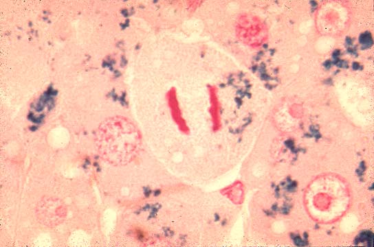
Mitoses |
Fig 63 - Mitoses in normal liver are very rare.They increase in regeneration after injury.The periportal hepatocytes have the highest regenerative activity being better nourished and oxygenated than the rest of the lobule.This slide shows a periportal hepatocyte in mitosis following partial hepatectomy, in the rat which was iron loaded.
|
|
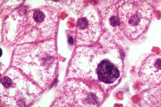
Binucleated hepatocytes |
Fig 64 - These binucleated cells increase in accelerated liver cell renewal following
an injury.Their presence, therefore, has a diagnostic significance.This slide shows many
binucleated hepatocytes in a liver with obvious lobular inflammation.The binucleation is the
result of nuclear division not followed by cytoplasmic division.
|
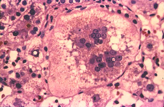
| Polyploid Nuclei |
Fig 64a - Presence of hepatocytes with many polyploid nuclei is another manifestation of increased liver cell renewal.Notice in this slide the nucleus of one hepatocyte being much larger than the rest.Polyploidy is the result of a mitosis that has completed the DNA replication phase but did not enter the phases of division.
|
|
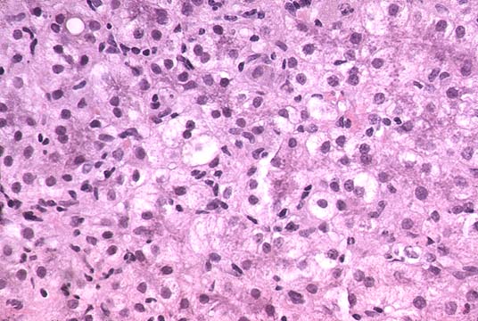
Multinucleation |
Fig 65 - In certain inflammatory and necrotic processes regenerating hepatocytes become highly multinucleated. This phenomenon is seen especialy in children. This slide shows a hepatocyte with more than 20 nuclei in a case of so-called "neonatal hepatitis".
|
The factor(s) stimulating proliferation of remaining hepatocytes are not known.Tey may be humoral. Indeed: 1)blood from a partially hepatectomized animal induces a proliferative response in the liver of a non- operated animal. 2)partial hepatectomy in one of two rats joined by carotid artery/jugular vein anastomosis induces proliferative response in the liver of the other rat.3)in heterotropic liver autografts supplying arterial blood through the hepatic vein instead of th portal vein, hepatocyte regeneration occurs around the central lobular vein instead of around portal tracts.
As in every other organ, exagerated regeneretion may induce hyperplastic changes (see the discussion on hyperplasia from table of contents).
CONTENTS | PIGMENTS