INFLAMATION


In the liver, as in every vascularized tissue, cell death (necrosis) evokes an inflammatory reaction which is morphologically manifested by the appearance of inflammatory cells together with edema and congestion around parenchymal cells which, in the case of the liver, are damaged hepatocytes or damaged bile ducts or damaged blood vessels. Therefore we talk of "HEPATITIS", "CHOLANGITIS", "VASCULITIS".

Inflammation of the liver lacks the gross classical signs of swelling (tumor), reddening (rubor), pain (dolor), but< microscopically it classically shows presence of inflammatory exudate with morphologically documentable cytological and tissue changes.

The type and distribution of the inflammatory exudate and the type of changes will vary sometimes according to the etiological agent. We say sometimes because, in the liver, the inflammatory reaction may not help to clearly distinguish between an hepatitis due to a chemical agent9r to an infectious agent such as a virus, e.g. it may not distinguish between a viral and a toxic hepatitis.

Obviously we encounter in the live acute and chronic inflammation.


INFLAMMATION: Lobular changes

Diffuse lobular inflammation

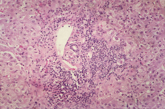
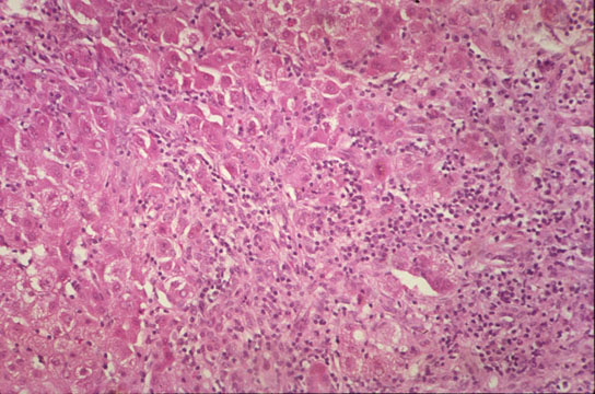
Fig 53 - DIFFUSE LOBULAR INFLAMMATION: Diffusely scattered mononuclear cell infiltration of the lobule especially in the perivvenular area. Lymphocytes and monocytes predominate but neutrophils, eosinophils and plasma cells may be present.

Kupffer cell hyperplasia

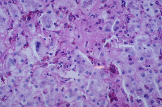
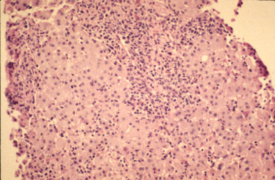
Fig 54 - KUPFFER CELL HYPERPLASIA: With cells full of lipofuscin an bile, result of phagocytosis of disrupted hepatocytes. Nodules of these cells may persist after healing (late nodules of Spatknoetchen).

Liver cell regeneration

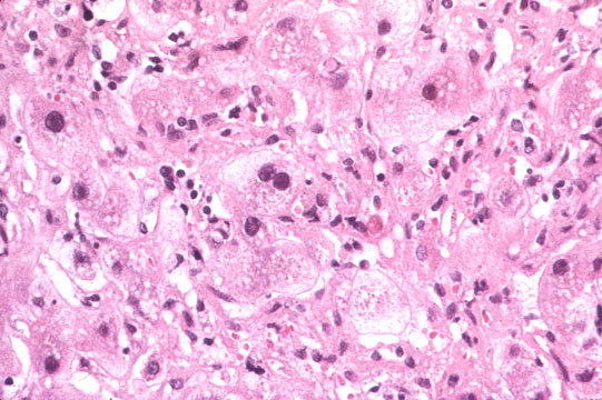
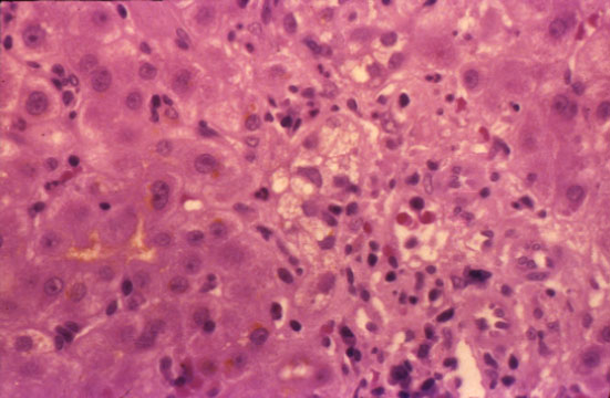
Fig 55 - LIVER CELL REGENERATION: Expressed by the presence of binucleate cells obviously due to increase rae of liver cell death. Mitoses are rare in the liver.

Central vein endophlebitis

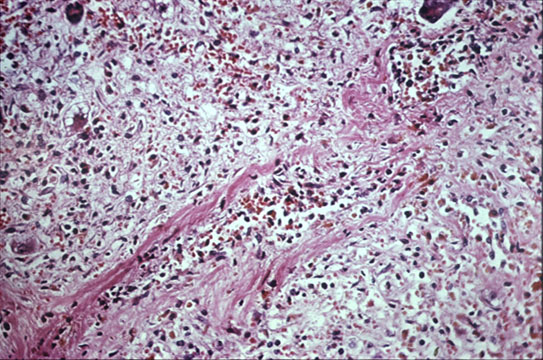
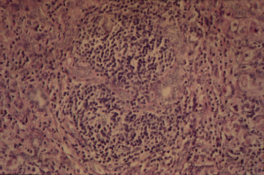
Fig 56 - CENTRAL VEIN ENDOPHLEBITIS: In a case of fulminatinaute viral hepatitis..

De-glycogenation

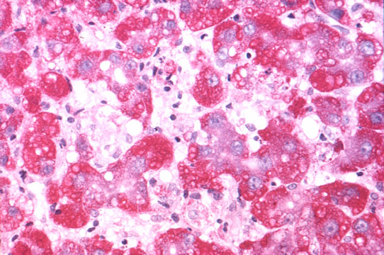
Fig 57 - DE-GLYCOGENATION: Injured cells lose their glycogen at very early stages. These same cells are those that release cytoplasmic enzymes in the serum. PAS stain. Glycogen stains red..

Intracellular and canalicular cholestasis

Fig 58 - INTRACELLULAR AND CANALICULAR CHOLESTASIS: May be present and very prominent i certaincdinsha in tscaefcostti orm of viral hepatitis A.


RETICULUM
Death of single of single or small groups of hepatocytes may leave the reticulum intact, cell regeneration will fill up the gaps and the damage will be completely repaired (ad integrum). In areas, however, where the reticulum is damaged, healing can occur only by scar and the process will lead to fibrosis. If, however, many scars are produced throughout the liver, the blood circulation may be affected and rearranged. The damage will lead to cirrhosis.


PORTAL AND PERIPORTAL INFLAMMATION
Portal inflammation may involve all or some portal fields. In acute viral hepatitis most portal fieds are infiltrated with inflammatory cells, mostly lymphocytes with some plasma cells containing IgG (1) and iron laden macrophages. Lymphocytic infiltration is sometimes present in the form of lymphoid follicles even with germinal centers and when it is associated with portal bile duct damage consisting in stratification, vacuolization and lymphocytic infiltration as in chronic active hepatitis C we call it hepatitic bile duct lesion Poulsen-Cristoffersen lesion) (Poulsen, Human Path, 3:217, 1972).

Portal inflammation may also be accompanied with bile extravasates, granulomas, purulent exudate, destruction of bile ducts, ductular reaction, fibrosis and may extravasate into the periportal liver parenchyma thus disrucpting the periportal limiting plate of hepatocytes. And from there, it may spread to the central acinar area and even reach an adjacent portal field to form a porto-portal septum. In this sequence a peculiar relationsh between inflammatory cells and hepatocytes develops at the parenchymal/mesenchymal interface such as periphery of portal fields and periphery of newly formed inflammatory septa. There is destruction of single or small groups of hepatocytes which are surrounded by and in close membrane contact with lymphocytes and macrophages. The phenomenon has been called:


"PIECEMEAL NECROSIS"
The surrounding and membrane contact between lymphocytes and hepatocyte which goes under the name of "PERIPOLESIS" is the hallmark of piecemeal necrosis. When lymphocytes penetrate a hepatocyte or any other cell, the phenomenon is called "EMPERIPOLESIS". In piecemeal necrosis the hepatocytes may become necrotic or apoptotic. Others may become hyperplastic and grow in "ROSETTES" surrounded by a rim of fibrous tissue. Ductular proliferation may occur. The portal field undergoes fibrous expansion.

Periportal and periseptal necrosis of hepatocyte without peripolesis should not be diagnosed as piecemeal necrosis which is the typical lesion of chronic active hepatitis and vacuolar degeneration of chronic cholestasis.

Piecemeal necrosis

Fig 59 - PIECEMEAL NECROSIS: In a patient with chronic active hepatitis B. Notice the disruption of the limiting periportal plate by dense mononuclear cell infiltrate which surrounds hepatocytes (peripolesis).

Lymphocytic spillover

Fig 60 - LYMPHOCYTIC SPILLOVER: In case of acute hepatitis A. There is no peripolesis nor rosetting fibrosis.

"Biliary" piecemeal necrosis

Fig 61 - "BILIARY" PIECEMEAL NECROSIS: In a case of "primary biliary cirrhosis". There are vacuolated pseudoxanthomatous cells and no peripolesis nor rosetting fibrosis.

Hepatitic bile duct lesion
(Poulsen Christoffersen lesion)

Fig 62 - HEPATITIC BILE DUCT LESION (Poulsen-Christoffersen Lesion): Lymphoid nodule surrounding a damaged portal bile duct.


CONTENTS