LIVER FIBROSIS


Chronic liver diseases lead to fibrosis which leads to derangement of the architecture, portal hypertension and may produce such an irreversible rearrangement of the circulation as to cause cirrhosis. There is a fine line between fibrosis and cirrhosis. Fibrosis is not only the result of necrosis, collapse and scar formation but also the result of derangements in the synthesis and degradation of matrix by injured mesenchymal cells that synthesize the various components of the matrix which in the liver are the following categories:


COLLAGENS
TYPE I COLLAGEN: the most abundant collagen of mature scar tissue present in portal fields and fibrous septa.
TYPE III COLLAGEN: Also abundant as type I and very abundant in distensible organs such as arteries and skin.
TYPE IV COLLAGEN: this type is present in the matrix surrounding epithelial cells, therefore in the liver is in the sinusoids and in the basement membranes of vessels and bile ducts.
TYPE V COLLAGEN: fine collagen fibrils which probably constitute the core of larger fibrils. It increases in cirrhosis where they surround hepatocytes.
TYPE VI COLLAGEN: it is found throughout the connective tissue and its function is probably that anchoring structures to collagen fibers.
TYPE VII COLLAGEN: the largest collagen apparently functions as an anchoring agent between basement membranes and underlying stroma. Collagens type II, VIII, IX, X, XI, XII, and XIII are not found in the liver.

GLYCOPROTEINS
LAMININ: is a glycoprotein that is distributed with collagen IV. It is increased in perisinusoidal fibrosis. It can be measured in the serum and correlates with the degree of fibrosis. Consists of 3 polypeptide chains.
FIBRONECTIN: consists of 2 polypeptide chains. It mediates adhesion of collagen, fibrin and heparin to cells, therefore it is involved in the organization of thrombi and in wound healing inducing attachment of these structures to cells.
ENTACTIN: is a polypeptide apparently a facilitator of laminin.
UNDULIN: A 3 chain polypeptide associated with types I and II collagen, thus present in portal fields. It might facilitate the cross linkage of these fibers.
ELASTIN: Present in the wall of blood vessels and in the capsule and increases in fibrosis.

PROTEOGLYCANS
Chondroitin sulphate, dermatan sulphate, keratan sulphate, heparan sulphate and heparin. They interact between cell surface receptors and macromolecules such as growth factors, collagens, fibronectin, laminin etc.

MATRIX PRODUCTION AND DEGRADATION
All categories of liver matrix are produced by hepatocytes, lipocytes which are in the space Disse, fibroblasts and myofibroblasts. Necrotic cells after injury evoke reaction of inflammatory cells which secrete mediators like cytokines which stimulate matrix producing cells. Fibrosis develops after repeated and persistent injury that overcomes the degrading ability of matrix on the part of the liver that attempts to eliminate those formations through degrading enzymes which are produced by fibroblasts, neutrophils and macrophages. They are: interstitial collagenases, stromelysin, Type IV collagenase-gelatinase.
EVALUATION OF HEPATIC FIBROSIS
HISTOLOGICAL: Masson trichrome stain. Siler reticulin stain. Specific antibodies for collagen types. Dsmin and Vimentin for lipocytes. Vimentin for myofibroblasts.
BIOCHEMICAL: Determination of various enzymes in matrix synthesis are of very limited usefulness. Serum laminin in benign fibrosis.


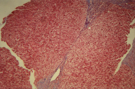
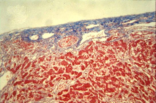
Central fibrosis, mild

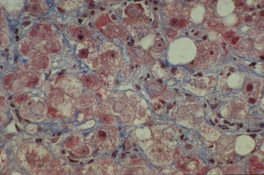
Fig.66 - CENTRAL FIBROSIS, MILD: The mild form consists of deposition of fine collagen fibers in the sinusoids of the pericentral lobular region. This form is called "chicken wire fibrosis".

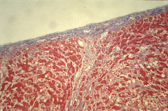
Central fibrosis, severe

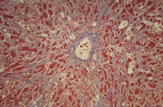
Fig.67 - CENTRAL FIBROSIS, SEVERE: This form of fibrosis mostly occuring in alcoholics goes under the name of "sclerosing hyalin necrosis". It consists of wide pericentral areas of fibrosis that may extend to portal fields.

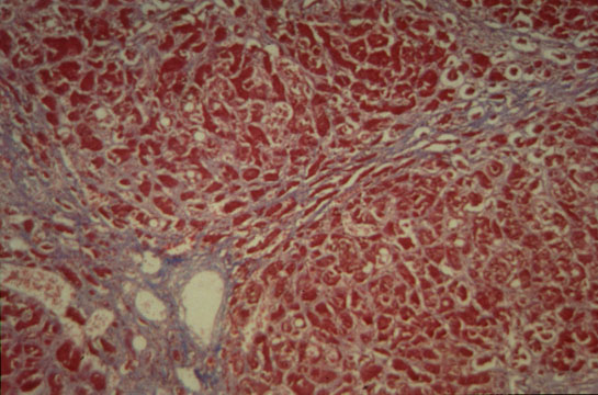
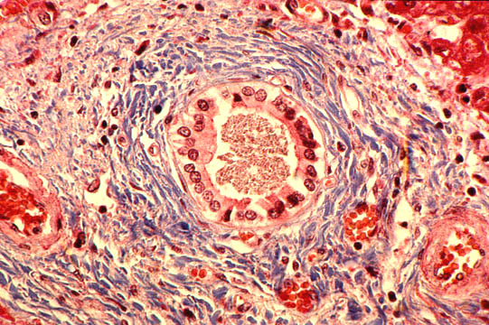
Portal and periportal fibrosis

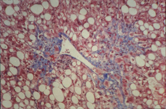
Fig.68 - PORTAL AND PERIPORTAL FIBROSIS: Fibrous expansion of portal fields with fibrosis extending along the terminal centracinar portal veins assuming the appearance of "stellate" fibrosis. Expansion and branching of normal portal fields must not be confused with pathological portal fibrosis which contains inflammatory cells.

Bridging fibrosis
Central-central

Fig.69 - BRIDGING FIBROSIS: Central-central. Connects central veins with central veins. It is rare and occuring mostly in chronic passive congestion as in this case.
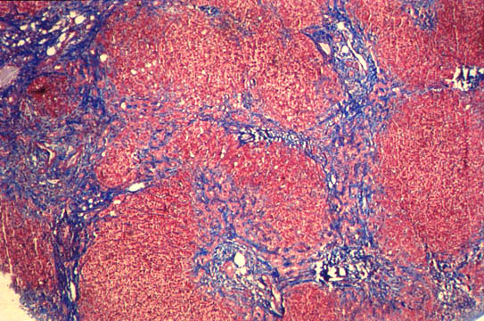
Bridging fibrosis
Porto-portal

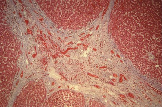
Fig.70 - BRIDGING FIBROSIS: Porto-portal. This form is common and usually associated with porto-central fibrosis. It follows a portal inflammation that extends to the terminal, centroacinar, portal venules.
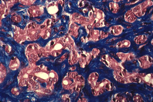
Bridging fibrosis
Porto-central

Fig.71 - BRIDGING FIBROSIS: Porto-central. It occurs after centrolobular necrosis and produces new vascular connections between portal fields and central veins which may lead to cirrhosis. It is the most severe form of bridging necrosis and fibrosis and is most of the times associated with porto-portal fibrosis.


BILIARY FIBROSIS
It is characterized by portal fibrosis with marked ductular proliferation extending to porto-portal septa and transformation of the liver parenchyma into sharp "garland" shaped nodules. It should not be confused with cirrhosis since this type of biliary fibrosis that is due to a chronic biliary obstruction may be reversible. The plexiform ductular proliferation in portal fields and septa is the hallmark of this lesion.
Biliary fibrosis

Fig.72 - BILIARY FIBROSIS: Severe nodular transformation of the liver by porto-portal fibrosis septa without inflammatory cells in clean obstructive jaundice.

Biliary fibrosis

Fig.73 - BILIARY FIBROSIS: Portal area with marked ductular proliferation and minimal inflammation in a case of chronic biliary obstruction. To be distinguished from cirrhosis.

Biliary fibrosis

Fig.74 - BILIARY FIBROSIS: Concentric peribiliary fibrosis with some inflammatory cells in a case of ascending cholangitis.


LIVER COLLAPSE
Collapse has to be differentiated from fibrosis. It is due to disappearance of hepatocytes and approximation of the remaining reticulin fibers. When it is focal produces connecting septa; when it involves large lobular areas and multiple lobules, it produces approximation of portal fields together which can still be recognized with a stain for elastic fibers. If the collapse is submassive it may have a favorable outcome. If it is massive, it will be fatal.
Capsular fibrosis

Fig.75 - CAPSULAR FIBROSIS: Thickening of the Glisson capsule that, when is focal, may be seen in normal liver.

Subcapsular fibrosis

Fig.75 - SUBCAPSULAR FIBROSIS: It is a normal structure due to arching of the subcapsular septa back to the capsule forming parenchymal nodules. These septa are only 2mm deep. The finding must not be interpreted as cirrhosis.


CONTENTS