|
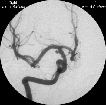
Digital Subtraction Angiogram
Identify the following structures on the anterior-posterior angiogram of the internal carotid artery:
Additional angiograms will be posted in the near future. |

| Neurovascular Anatomy: Internal Carotid Artery: Angiogram |
|
Digital Subtraction Angiogram
Identify the following structures on the anterior-posterior angiogram of the internal carotid artery:
Additional angiograms will be posted in the near future. |
|
|
|
INTERNAL
CAROTID AND BRANCHES |
|
Move
the cursor along the course of the vessels on the angiogram to
identify individual segments.
|
|
A1
Segment |
|
From Internal Carotid Bifurcation to Anterior Communicating Artery. NEUROVASCULAR
SYNDROME Penetrating
Branches:
|
|
Anterior
Communicating Artery
|
|
Connects
bilateral anterior circulations. |
|
Recurrent
Artery of Heubner
|
|
Supplies
head of caudate and anteroinferior internal capsule. |
|
Pericallosal
Artery
|
|
Continuation
of the Anterior Cerebral Artery as it arches superiorly and posteriorly. |
|
Horizontal
(M1) Segment
Middle Cerebral Artery |
|
Branches include lateral lentciulostriate arteries. NEUROVASCULAR
SYNDROME SENSORY
|
|
Sylvian
(M2) Segment
Middle Cerebral Artery |
|
Segment
divides into superior and inferior divisions which can be a site
for an embolus to lodge. Inferior
Division Infarction: |
|
Cortical
(M3) Segment
Middle Cerebral Artery |
|
Distal branches of MCA course laterally to insular cortex and loop around operculum - "Candelabra" effect seen on lateral angiograms. Embolization
of individual cortical branches can produce highly circumscribed
infarctions accompanied by specific neurologic deficits. |
|
Lateral
Lenticulostriate Arteries |
|
Branch of M1 Segment of MCA. Supplies basal ganglia structures: Part of head and body of caudate, globus pallidus, putamen, and the posterior limb of the internal capsule. NEUROVASCULAR
SYNDROME |
|
Supraclinoid
Segment
Internal Carotid Artery |
|
Begins after penetration of dura, continues until bifurcation into Anterior and Middle Cerebral Arteries Three
Branches: |
|
Cavernous
Segment
Internal Carotid Artery |
|
Passes through cavernous sinus with Abducens Nerve. Branches
supply posterior pituitary (Meningohypophyseal Artery). |
|
Petrous
Segment
Internal Carotid Artery |
|
Extends
from base of skull to apex of petrous bone
Enters cranial vault via foramen lacerum. Branches normally not seen angiographically - may be enlarged with carotid occlusive disease. |
|
Cervical
Segment
Internal Carotid Artery |
|
Begins
at the bifurcation of Common Carotid Artery (level of C4). |
|
Posterior
Communicating Artery
|
|
Second branch of supraclinoid internal carotid. Connects
Supplies
thalamus, hypothalamus, optic chiasm, and mamillary bodies.
Common
site for aneurysms -
AS IS SEEN IN THIS CASE |
|
Ophthalmic
Artery
|
|
Usually
arises intradurally (80-90%), below anterior clinoid process.
Supplies
globe, orbit, frontal scalp, the frontal and ethmoidal sinuses.
Ophthalmic
artery branches anastamose with Maxillary artery branches - potential
for collateral flow in cases of proximal carotid occulsion.
|
|
Anterior
Choroidal Artery
|
|
Last branch to originate from Internal Carotid Artery. Cisternal
segment: Artery passes through crural cistern, supplies optic
tract, posterior limb of internal capsule, branches to midbrain,and
lateral geniculate nucleus.
Plexal
segment: Supplies choroid plexus of anterior portion of temporal
horn of lateral ventricles.
|
|
Anterior
Cerebral Artery
|
|
One of the terminal branches of internal carotid artery. A1 Segment:
A1 Branches:
Infarction
syndrome:
|