This is the current version on airways 06/06/00
Read first the text book: Computed Tomography and Magnetic resonance of the Thorax by Nadich et all
Then go through this exercise to assess your comprehension
This lesson is on recognition of normal airways. You need to have a clear understanding of Anatomy of bronchial tree especially three dimensional perspective. Recognition of airways by CXR, Bronchograms and CT will be reviewed.
Q: Describe extent and relationships of Trachea.
Larynx (C6) to Carina (T5)
Slight deviation to right
Walls are parallel
Courses antero-posterior obliquely, hence the position will vary depending on the level of sections
Progressive caudal sections will show trachea more posteriorly
Lies anterior and slightly to right of Esophagus
In contact with mediastinal pleural reflection of RUL (right Para tracheal stripe)
Potential space between trachea and esophagus and is occupied by lungs (retro tracheal recess) (posterior tracheal band in lateral chest)
Q: Describe the appearance and dimensions of Trachea .
Walls are parallel
Thin wall , well defined internally by air and externally by fat
Smoothly serrated contour
Horse shoe shaped cartilage rings at regular intervals
Flat membranous part posteriorly
Variations in shape and size common
Cross sectional appearance varies from rounded, oval to horseshoe shaped with a flatter posterior wall
Mean AP diameter 20 mm
Mean Transverse diameter 17 mm
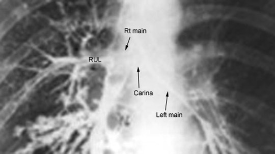
Q: What is the location and the mean angle of carina?
At the level of T5 and louis angle of sternum, trachea (carina) divides into left and right main stem bronchi
Mean carinal angle is 56 degrees
Q: What is the length diameter and direction of right maim bronchus ?
More straight and in line with Trachea in adults, reason for aspiration being more common on right
Symmetrical to left upto 15 years, reason why aspiration is equal on both sides at this age.
Transverse diameter of right main bronchus is 15 mm
2.2 cm long
Q: What is the length diameter and direction of left maim bronchus
Transverse diameter of left main bronchus is 13 mm
5 cm long
Q: What is the origin, length, diameter, direction and divisions of right upper lobe bronchus?
Arises from lateral aspect of right main bronchus
2.5 cm from carina
Horizontal orientation
Seen as round lucency on lateral (end on view)
Divides into anterior, posterior and apical segments
Anterior and posterior segments have horizontal orientation
Apical segment has a vertical orientation
Q: What is the origin, length, diameter, direction and divisions of right intermediate bronchus
From RUL to RML and RLL
3-4 cms long
Has a vertical orientation
Q: What is the origin, length, diameter, direction and divisions of right middle lobe
Arises from anterolateral aspect of intermediate bronchus
Opposite the origin of superior segment of RLL
Has a horizontal orientation
Bifurcates into lateral and medial segments
Both of them have a horizontal orientation
Q: What is the origin, length, diameter, direction and divisions of right Lower lobe
Superior segmental bronchus takes off opposite RML along the posterior aspect of RLL bronchus
Superior segement has a horizontal orientation
Medial basal segment takes off along medial side of RLL bronchus
In AP projection basal segments are in the following order anterior lateral, posterior and medial basal segments
In lateral same relationship anterior - lateral - posterior (ALP)
Oriented in
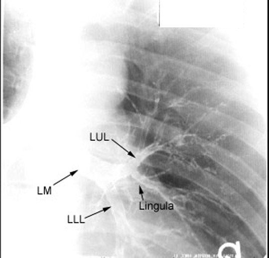
Q: What is the origin, length, diameter, direction and divisions of left maim bronchus
Transverse diameter of left main bronchus is 13 mm
5 cm long
Oriented obliquely
Q: What is the origin, length, diameter, direction and divisions of left upper division
Divides into LUL (upper division) and Lingula (lower division)
Horizontal orientation
LUL
1 cm long
Horizontal
Divides into apical-posterior and anterior segments
Apical-posterior segment has a vertical orientation
Anterior segemnt is oriented
Lingula
2-3 cm long
Oblique course
Divides into superior and inferior segments
Segments are obliquely oriented
Q: What is the origin, length, diameter, direction and divisions of left lower lobe
Superior segment takes off posteriorly first
Superior segment is oriented horizontally
No separate medial basal segment
LLL beyond superior segment is vertically oriented
Same ALP relationship as right
Medial basal segment arises with anterior basal segment (anteromedial basal segment)
Q; List the tracheal stripes, normal size and significance of abnormality.
Right Para tracheal stripe width ranges from 1-4 mm
Width greater than 4 mm indicates disease
Posterior tracheal band seen on lateral
obliterated by cancer esophagus or disease of medial portion of RUL
Posterior wall of intermediate bronchus in line with trachea beyond RUL orifice
On left posterior wall of left main does not form a line because it is in contact with airless mediastinum and left pulmonary artery.
Q: What are the lucencies seen in lateral view over tracheal air column?
Two circular translucencies in line with tracheal air column is RUL and LUL
Let us go through concepts for recognition of airways by CT chest
Q: List Central airways
Trachea
Carina
Main-stem bronchi
Lobar
Sub segmental bronchi
Q: Central airways are easily definable with CT. How are they useful in reading the film?
Bronchi are important sites for disease
Provide road map into the lung parenchyma
Point of orientation for interpretation of hilum (Bronchi serve as a lattice on which pulmonary arteries and veins drape)
Ability to visualize Bronchial anatomy depends on
Size
Orientation
Q: List Bronchi with horizontal orientation
RUL
Anterior segment
Posterior segment
LUL
Anterior segment
Middle lobe
Medial segment
Lateral segment
Superior segmental bronchus of Lower lobes
Q: Bronchi with vertical course (Circular lucencies)
Apical segment of RUL
Apical-posterior segment of LUL
Bronchus intermedius
Proximal portion of both lower lobes beyond superior segmental bronchus
Q: Bronchi with oblique course (difficult to visualize) (oval or elliptical in shape)
Lingular bronchus
Superior segment
Inferior segment
Lateral and medial segments of middle lobe
Q: Is visualization of bronchi in the periphery normal?
Visualization of bronchi in the periphery of lung is abnormal suggesting thickening of wall or parenchymal disease
Now we are ready to review CT sections.
For easier review I am going to take you through right bronchial tree and left bronchial tree in sequence.
Q: What are the CT sections by whch you evaluate the right lung bronchial segmental anatomy
Five characteristic sections
Distal trachea carina
RUL bronchus
Bronchus intermedius
Middle lobe bronchus
Lower lobe bronchus
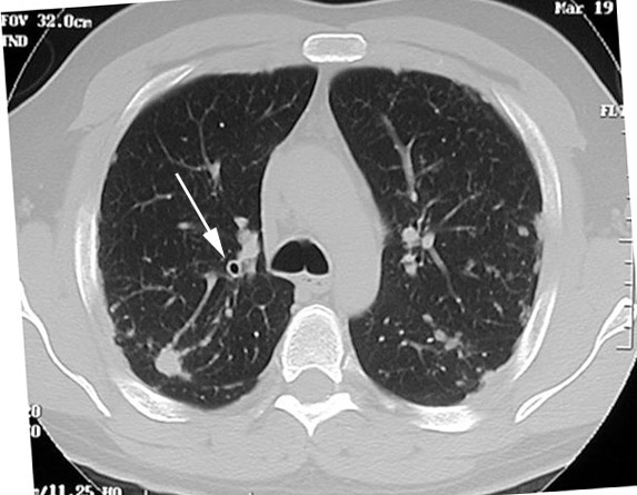
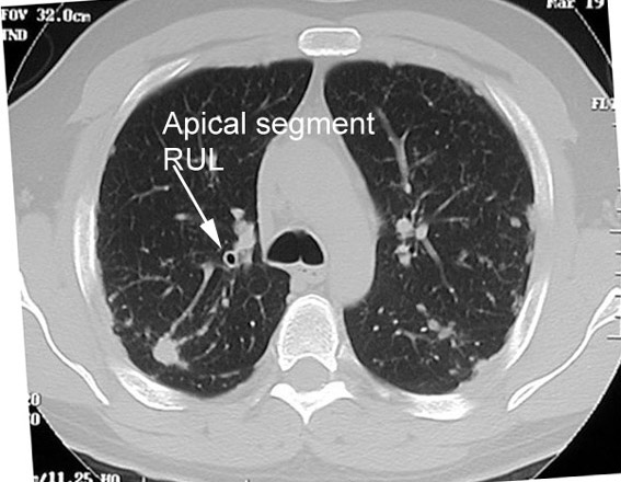
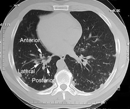
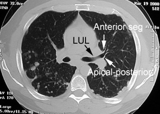
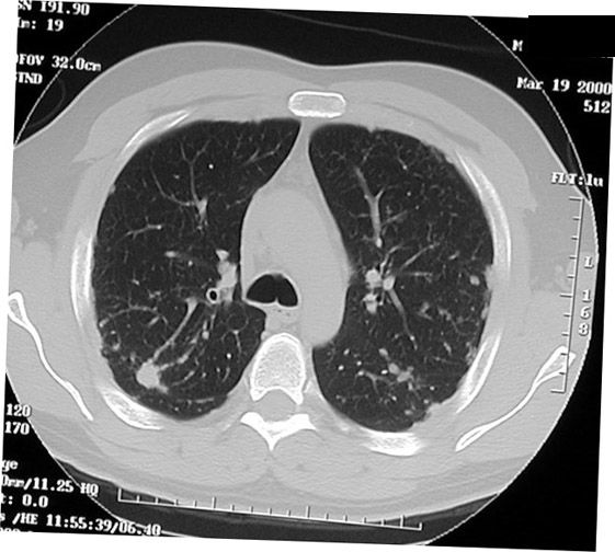
Q: Click to look at the section at the level of Carina. Identify the bronchial segment the arrow is poiniting to.
Answer
Apical segmental bronchus of RUL
Since the apical segment of RUL is vertical, section at this level gives a circular lucency.
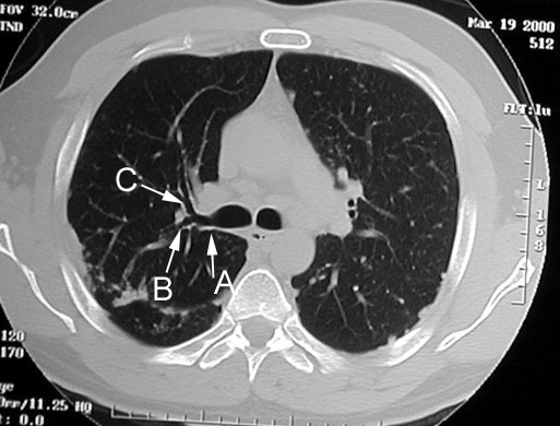
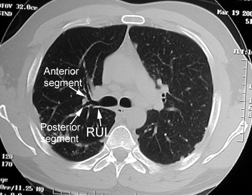
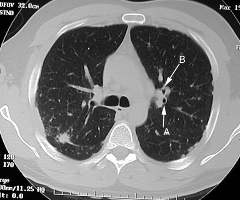
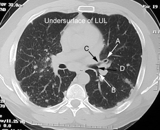
Q: You are looking at section at the level of RUL bronchus (at or just below carina). Identify the labeled airways.
Answer
They appear tubular in this section, since all of them course horizontally
Origin of apical segment of upper lobe superimposed on the distal portion of RUL as rounded decreased density not evident in this section.
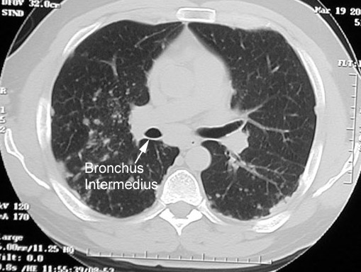
Q: You are looking at section at the level of bronchus intermedius.
What is the extent of bronchus intermedius?
Answer
Circular lucency since it has a vertical orientation.
Extends from RUL bronchus to RML bronchus
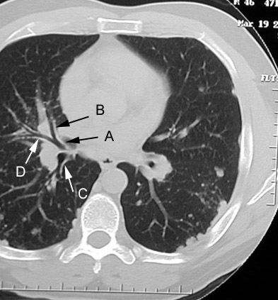
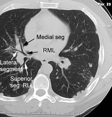
Q: You are looking at section at the level of RML bronchus Identify the labeled airways.
Answer
Middle lobe bronchus origin also marks beginning of right lower lobe
Extends anteriorly at a slightly oblique angle
Divides into medial and lateral segments
Since RML extends inferiorly the medial and lateral segments may be located in a lower plane
Triangular spur at the site of bifurcation
Superior segment of RLL arises at the same level as RML or at a slightly lower level
Superior segment of RLL courses posteriorly and runs in a horizontal plane
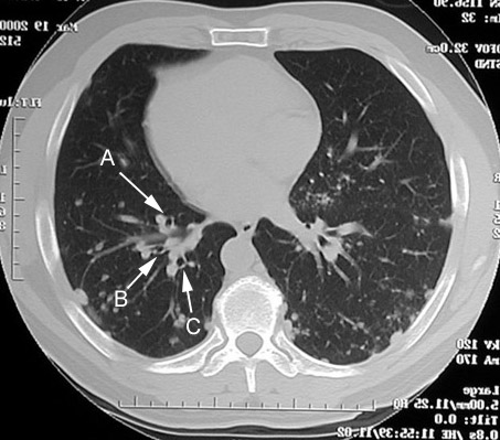
Q: You are looking at section at the levell of RLL basilar segments Identify the labeled airways.
Answer
Lower lobe beyond superior segment is vertical and is recognized as a circular lucency
Lies medial and anterior to lower lobe pulmonary artery
Appears suspended by superior portion of the inferior pulmonary ligament
Medial basilar segment arises first, anterior to inferior pulmonary vein
Anterior , lateral and posterior basilar bronchi course towards the anticipated positions
Q: What are the CT section by whch you evaluate the left lung bronchial segmental anatomy ?
Lower trachea and carina
Upper portion of LUL
Lower portion of LUL
Upper lobe spur
LLL basilar segments
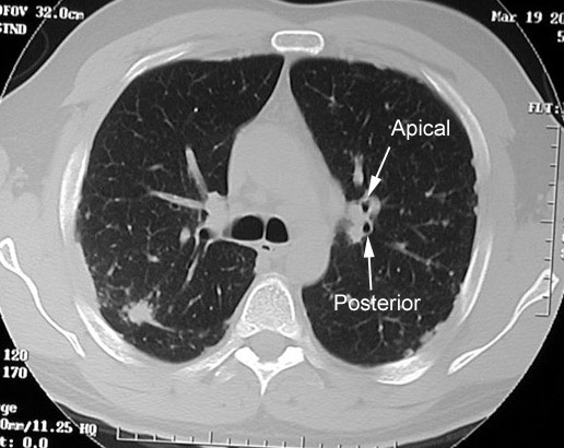
Q: You are looking at section at the level of lower trachea and carina. Identify the labeled airways.
Answer
Apical-posterior segmental bronchus LUL
Apical segment
Posterior segment
Seen as circular lucencies.
Apical-posterior segmental bronchus is separated from the left main bronchus by the left main pulmonary artery, which courses over LUL bronchus at this level
Q: Left upper lobe
LUL bronchus originates lower than RUL bronchus
Forms a sling over which the main pulmonary artery passes
The LUL bronchus is large
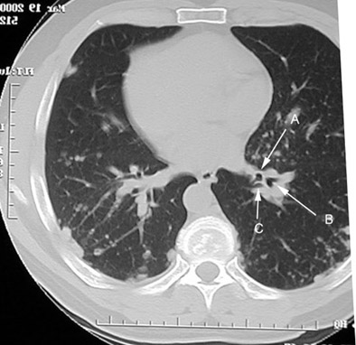
Q: You are looking at section at the level of upper portion of LUL Identify the labeled airways.
Answer
Posterior wall is smooth and slightly concave caused by the left pulmonary artery
The origin of the apical-posterior segmental bronchus can be recognized as area of increased lucency on the distal portion of LUL
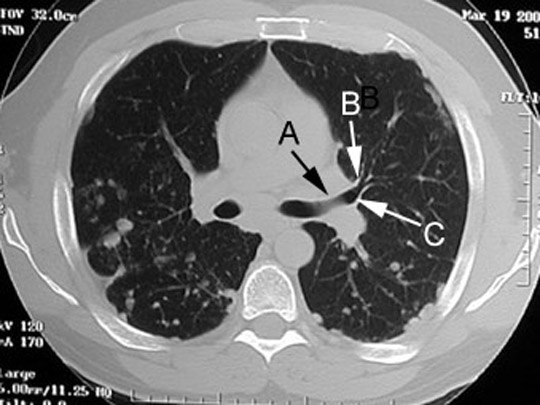
Q: You are looking at section at the level of lower portion of LUL Identify the labeled structures.
Answer
Left upper lobe spur is the landmark Triangular in shape.
Spur marks the origin of LLL (secondary carina)
Superior segment of LLL runs horizontally
The lingular bronchus arises from the undersurface of the distal portion of LUL
Courses obliquely inferiorly
Can be recognized by increased radiolucency at the distal portion of LUL similar to apical-posterior segment
Q: Section below LUL bronchus
Lingular bronchus is oval or elliptical separated spatially from LLL bronchus
Superior and inferior segments originate at a considerable distance from origin of lingular bronchus and often not recognized
Anterior segment of LUL is the only segment coursing anteriorly in a horizontal plane
Highly variable in origin from LUL
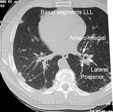
Q: You are looking at section at the level of LLL basilar segments Identify the labeled airways.
Answer
Same as right
Superior segment of LLL is similar to right side
Proximal portion of LLL below the superior segment is suspended by the inferior pulmonary ligament
Basal segments of LLL are similar to right side
Medial and anterior basal segments arise as a common trunk
Now let us concentrate on relationship of airways to other structures.
Q: Click to look at the section at the level of Carina. Identify the labeled structures.
Identify the vessels adjacent to apical segment of RUL.
Answer
Labeled Image
RUL pulmonary artery. Artery lies just medial to bronchus
Right superior pulmonary vein. Vein just lateral to bronchus
Q: You are looking at section at the level of RUL bronchus (at or just below carina). Identify the labeled structures.
Identify the labeled vessels adjacent to RUL.
Answer
Labeled Image
Truncus anterior first major branch of right pulmonary artery lies anterior to RUL bronchus
Branch of right superior pulmonary vein lies at the angle between anterior and posterior segmental bronchi
Anterior and medial to truncus anterior lies RUL vein (the apical-anterior vein)
Q: You are looking at section at the level of bronchus intermedius. Identify the labeled structures.
Answer
Labeled Image
Extends from RUL bronchus to RML bronchus
Lies directly behind right main pulmonary artery
Medial to right intralobar pulmonary artery
Posterior wall in contact with superior segment of RLL
Azygoesophageal recess is along posteromedially separated by lung tissue
Inferior to bronchus right superior pulmonary vein lies alongside the lateral border of right interlobar pulmonary artery
Multiple veins can be identified in this location
Right main pulmonary artery at the lateral border of intermedius bronchus turns inferiorly and becomes interlobar pulmonary artery.
At this position the artery may have a triangular configuration
Q: You are looking at section at the level of RML bronchus Identify the labeled structures.
Answer
Labeled Image
Interlobar pulmonary artery is vertical and lies immediately lateral to RML and RLL bronchi
Artery has an oval configuration at this point
Right superior pulmonary vein lies medial to RBL bronchus and can be seen entering the upper portion of left atrium
Q: You are looking at section at the levell of RLL basilar segments Identify the labeled structures.
Answer
Labeled Image
Lower lobe beyond superior segment is vertical and is recognized as a circular lucency
Lies medial and anterior to lower lobe pulmonary artery
Appears suspended by superior portion of the inferior pulmonary ligament
Medial basilar segment arises first, anterior to inferior pulmonary vein
Anterior , lateral and posterior basilar bronchi course towards the anticipated positions
Branches of right interlobar pulmonary artery are round and lay posterolateral to the proximal portions of basilar segments
Inferior pulmonary veins are oriented horizontally and can be traced to right inferior pulmonary vein and subsequently into the lower portion of left atrium.
Q: What are the CT section by whch you evaluate the left lung bronchial segmental anatomy ?
Lower trachea and carina
Upper portion of LUL
Lower portion of LUL
Upper lobe spur
LLL basilar segments
Q: You are looking at section at the level of lower trachea and carina. Identify the labeled structures.
Answer
Labeled Image
Apical-posterior segmental bronchus is separated from the left main bronchus by the left main pulmonary artery, which courses over LUL bronchus at this level
LUL pulmonary artery cab be traced to its origin from the left main pulmonary artery and lies posterolateral to the left superior pulmonary vein
Superior pulmonary vein tends to be located anteriorly
Q: Left upper lobe
LUL bronchus originates lower than RUL bronchus
Forms a sling over which the main pulmonary artery passes
The LUL bronchus is large
Q: You are looking at section at the level of upper portion of LUL Identify the labeled structures.
Answer
Labeled Image
Posterior wall is smooth and slightly concave caused by the left pulmonary artery
Superior segment of Lower lobe abuts the posterior portion
Posterior wall of LUL bronchus is slightly convex because it is indented superiorly and posteriorly by the left main pulmonary artery
Posterior to LUL bronchus the left pulmonary artery continues as interlobar pulmonary artery
Appears triangular at this level because of the change in course
Left superior pulmonary vein lies in front of LUL bronchus and has a horizontal course
Distinct branching can be identified peripherally
Q: You are looking at section at the level of lower portion of LUL Identify the labeled structures.
Answer
Labeled Image
Left upper lobe spur is the landmark Triangular in shape.
Spur marks the origin of LLL (secondary carina)
Left interlobar artery has a vertical course and lies lateral to the spur
Lateral to the origin of the lingular bronchus the pulmonary artery to lingula can be identified
Directly anterior to LUL bronchus is the inferior portion of the left superior pulmonary vein
Pulmonary parenchyma can be seen between pulmonary artery and descending aorta (retro bronchial stripe)
Q: You are looking at section at the level of LLL basilar segments Identify the labeled structures.
Answer
Labeled Image
Proximal portion of LLL below the superior segment is suspended by the inferior pulmonary ligament
Branches of pulmonary artery to the lower lobe lie lateral and posterior to the basilar bronchi
Left inferior pulmonary vein is horizontal and cab traced to left atrium
Left inferior pulmonary vein course along the lateral border of descending aorta
The session is over