The images are not necessarily from this case.
Normal stools
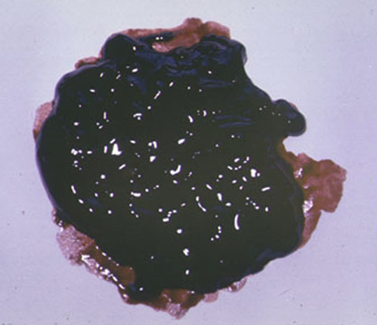
Black Stools Melena: the specimen consists of a black, tarry stool passed per anus. Note the mahogany color at the edge of the specimen (filter paper). Dr Ralph Leischner
Telengiectasia osler-weber-rendu syndrome/Clinical photo Dr Ralph Leischner
"spider nevus" alcholic cirrhosis Dr Ralph Leischner
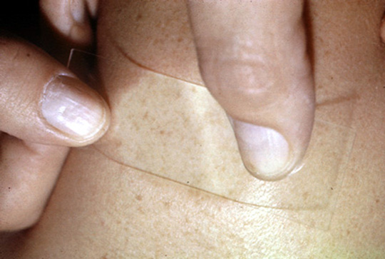
"spider nervus" compressed Dr Ralph Leischner
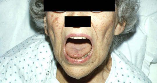
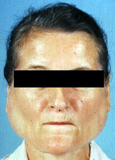
Parotid gland enlargement Sjogren's syndomre Dr Harry Messmore
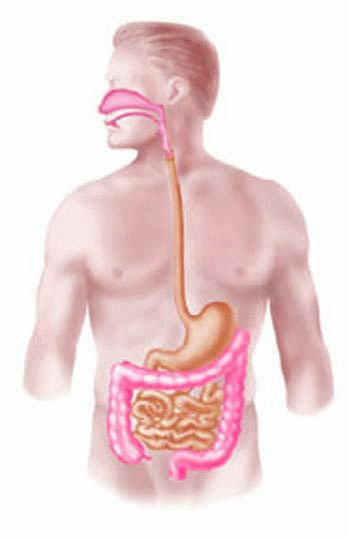
Upper GI tract / Ligament of Treitz
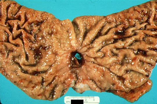
Duodenal ulcer The surgical specimen (opened) of the distal stomach and proximal duodenum reveals an irregular ulcer of the duodenal mucosa. The ulcer is filled with clotted blood. The duodenal mucosa is stained with blood. Dr Ralph Leischner
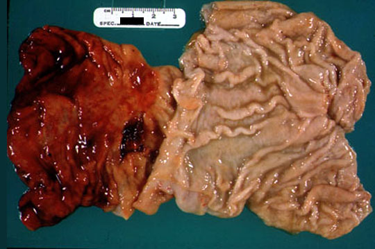
Gastric ulcer The surgical specimen of the distal stomach reveals a perforated, peptic ulcer. Dr Ralph Leischner
Hemorrhagic gastritis: the stomach is opened revealing diffuse hyperemia of the gastric mucosa. Dr Ralph Leischner
Esophagitis
Esophgeal varices
Mallory-weiss tear
AV malformation
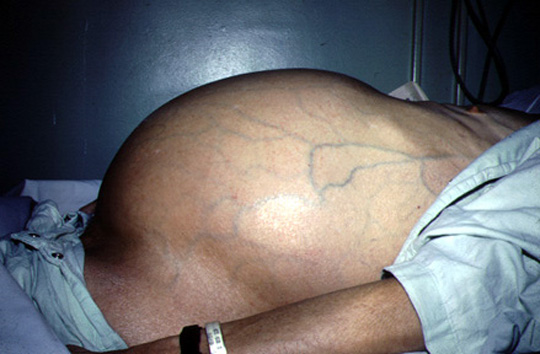
Ascites Dr Ralph Leischner
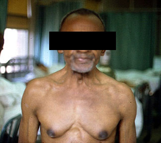
Hyperestrogenism Gynaecomastia sec to estrogen, dark areola. Dr Arcot Chandrasekhar
Coffee grounds
Red blood and clots
Bile stained
Clear
EGD
Pale conjunctiva
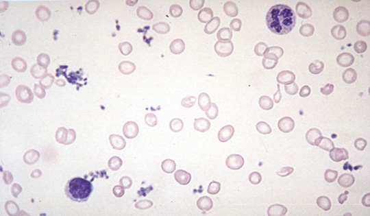
Hypochromic microcytic anemia Dr Ralph Leischner
Hemo-occult positive stools
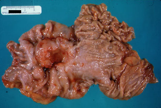
Cancer colon The surgical specimen consisting of terminal ileum and proximal colon reveals a large, exophytic adenocarcinoma in the cecum. Dr Ralph Leischner
Cancer stomach The surgical specimen of the distal stomach reveals an adenocarcinoma. The neoplasm contains a central, necrotic ulcer. The edges of the ulcerated neoplasm are elevated above the mucosal surface. Dr Ralph Leischner
Cancer small bowel
Hemorrhagic telengiectasia
Liver metastasis Dr Arcot Chandrasekhar
Liver CT normal
Liver Metastases
Send comments to Dr. A.J. Chandrasekhar M.D.