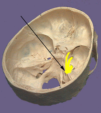
CN V. Trigeminal Nerve
The trigeminal nerve as the name indicates is composed of three large branches. They are the ophthalmic (V1, sensory), maxillary (V2, sensory) and mandibular (V3, motor and sensory) branches. The large sensory root and smaller motor root leave the brainstem at the midlateral surface of pons. The sensory root terminates in the largest of the cranial nerve nuclei which extends from the pons all the way down into the second cervical level of the spinal cord. The sensory root joins the trigeminal or semilunar ganglion between the layers of the dura mater in a depression on the floor of the middle crania fossa. This depression is the location of the so called Meckle's cave. The motor root originates from cells located in the masticator motor nucleus of trigeminal nerve located in the midpons of the brainstem. The motor root passes through the trigeminal ganglion and combines with the corresponding sensory root to become the mandibular nerve. It is distributed to the muscles of mastication, the mylohyoid muscle and the anterior belly of the digastric. The mandibular nerve also innervates the tensor veli palatini and tensor tympani muscles. The three sensory branches of the trigeminal nerve emanate from the ganglia to form the three branches of the trigeminal nerve. The ophthalmic and maxillary branches travel in the wall of the cavernous sinus just prior to leaving the cranium. The ophthalmic branch travels through the superior orbital fissure and passes through the orbit to reach the skin of the forehead and top of the head. The maxillary nerve enters the cranium through the foramen rotundum via the pterygopalatine fossa. Its sensory branches reach the pterygopalatine fossa via the inferior orbital fissure (face, cheek and upper teeth) and pterygopalatine canal (soft and hard palate, nasal cavity and pharynx). There are also meningeal sensory branches that enter the trigeminal ganglion within the cranium. The sensory part of the mandibular nerve is composed of branches that carry general sensory information from the mucous membranes of the mouth and cheek, anterior two-thirds of the tongue, lower teeth, skin of the lower jaw, side of the head and scalp and meninges of the anterior and middle cranial fossae.


