|
|
|
Move
the cursor along the course of the vasculature structures above
to identify individual segments and their perfusion targets. |
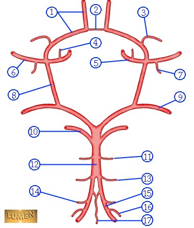
| Neurovascular Anatomy: Circle of Willis |
|
Circle of Willis
Identify the following structures on the left that comprise the circle of Willis:
Also try and identify the following:
|
|
|
|
Move
the cursor along the course of the vasculature structures above
to identify individual segments and their perfusion targets. |
|
A1
Segment |
|
From Internal Carotid Bifurcation to Anterior Communicating Artery. A1 Branches: NEUROVASCULAR
SYNDROME Penetrating
Branches:
|
|
Anterior
Communicating Artery
|
|
Connects
bilateral anterior circulations. |
|
Recurrent
Artery of Heubner
|
|
Supplies
head of caudate and anteroinferior internal capsule. |
|
Pericallosal
Artery
|
|
Continuation
of the Anterior Cerebral Artery as it arches superiorly and posteriorly. |
|
Horizontal
(M1) Segment
Middle Cerebral Artery |
|
Branches include lateral lentciulostriate arteries. NEUROVASCULAR
SYNDROME SENSORY
|
|
Sylvian
(M2) Segment
Middle Cerebral Artery |
|
Segment divides into superior and inferior divisions which can be a site for an embolus to lodge. Branches
supply: Inferior
Division Infarction: |
|
Cortical
(M3) Segment
Middle Cerebral Artery |
|
Distal branches of MCA course laterally to insular cortex and loop around operculum - "Candelabra" effect seen on lateral angiograms. Embolization
of individual cortical branches can produce highly circumscribed
infarctions accompanied by specific neurologic deficits. |
|
Lateral
Lenticulostriate Arteries |
|
Branch of M1 Segment of MCA. Supplies basal ganglia structures: Part of head and body of caudate, globus pallidus, putamen, and the posterior limb of the internal capsule. NEUROVASCULAR
SYNDROME |