|
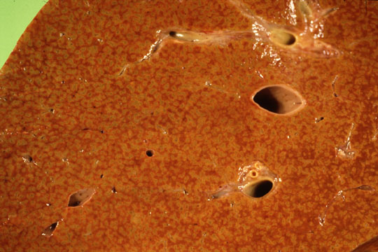
| Fig 1 - The cut surface of the liver reveals to the naked eye small,1.5 to 2 mm circular structures with a darker dot in the center and 4-5 darker dots at the periphery. These are the liver lobules. Notice a large radical of the hepatic vein and a large branch of the portal vein with adjacent bile duct. |
Portal Spaces of Lobule |
Fig 2 - Under light microscopy,the lobule has a roughly polygonal shape.The central dark dot is the central vein and the peripharal darker areas are the 4-5 portal spaces at the angles of the polygon. |
of Liver Lobule |
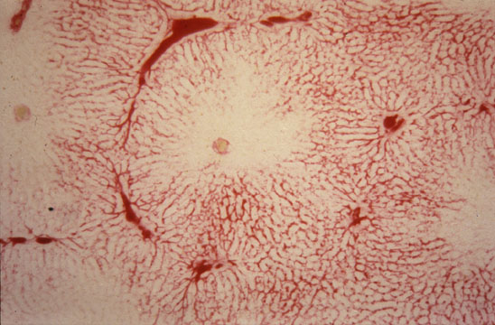
Fig 3 - Liver lobule demonstrated by portal microcirculation.. A dye was injected in the portal vein. Notice 6 portal areas around the lobule,the lightly injected central vein and lobular sinusoids and the terminal portal vein extending from the portal area and around the lobule.. The terminal portal vein area is not visible in an H&E section of a normal liver but becomes evident in cases of periportal infalammation and fibrosis. |