Differential Diagnosis: Ureteral Calculus
| Ureteral obstruction mimicking appendicitis. 18 year old male who presented with a two day history of right lower quadrant pain and vomiting. CT, shown below, was requested to evaluate for appendicitis. |
|
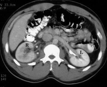
Figure 1. Contrast-enhanced CT image at the level of the kidneys shows delay in the right (R) nephrogram when compared to the left (L). Also note infiltration of the right perinephric fat (*). |
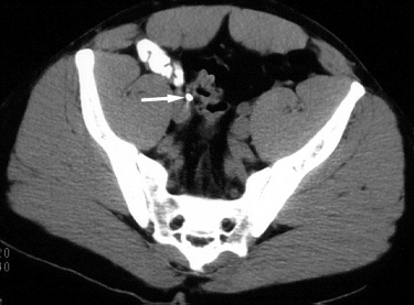 |
Figure 2. Contrast-enhanced CT image at the level of the midpelvis reveals a radioopaque calculus (arrow) in the right ureter. |
| The patient was given oral analgesic medications and discharged home from the emergency department. He spontaneously passed the calculus in his urine two days later and had relief of his symptoms. |
Return to Differential Diagnosis