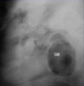
Plain film
Gall stone in a patient with ankylosing spondylitis
|
Plain filmGall stone in a patient with ankylosing spondylitis
|
|
Emphysematous cholecystitisAir in the lumen of Gall bladder |
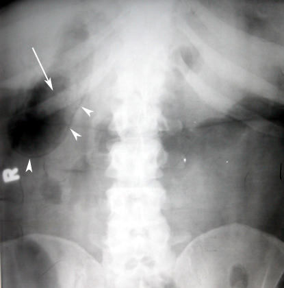 |
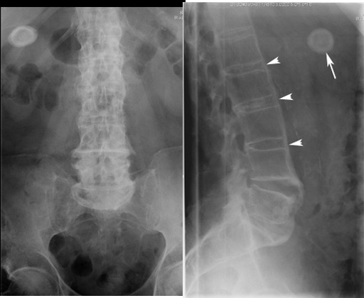
Emphysematous CholecystitisRadiograph showing:
|
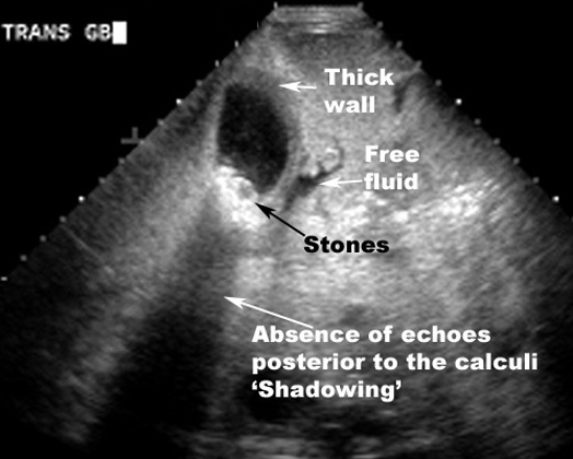
Ultrasound findings:
- Thick gallbladder wall - greater than 3 mm
- Stones present in gallbladder
- Pericholecystic fluid
- Sonographic Murphy's sign - tenderness over the gallbladder from the ultrasound transducer
- Emphysematous cholecystitis:
- Air in gallbladder
- Air in wall of gallbladder
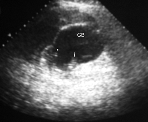
- Acalculus cholecystitis:
- Thickened gallbladder wall
- No stones seen
 |
Gallstones - Acute cholecystitissUS findings:
|
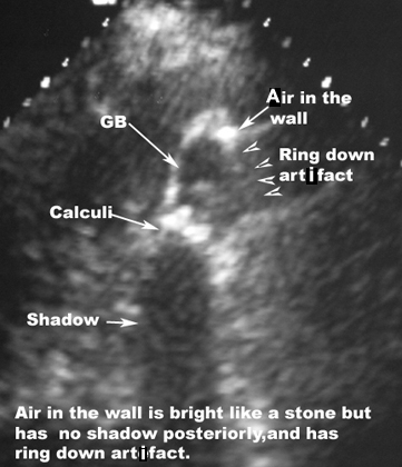 |
Acute Emphysematous CholecystitisUltrasound showing:
|
 |
Emphysematous Cholecystitis
Arrows point to ring down artifacts from air in the wall and lumen of GB.
|
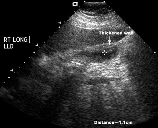 |
Acute acalculous CholecystitisUS Finding: Thickened gallbladder wall
|
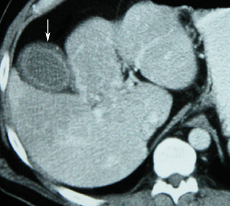 |
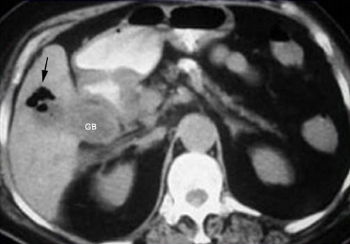
Acute CholecystitisCT showing:
|
|
Emphysematous cholecystitis |
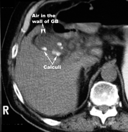 |
Emphysematous CholecystitisCT Findings:
|