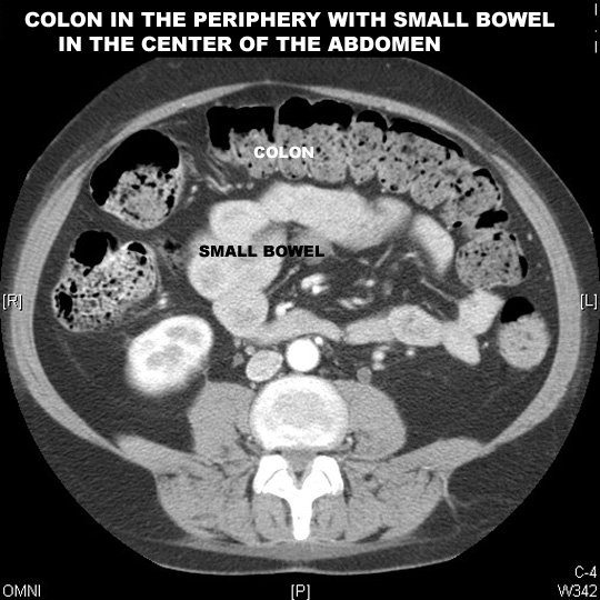
Bowel obstruction / Ileus

What are the sites of bowel obstruction?
What is ileus?

Dilated bowel which may be from:

What are the common causes of ileus?
What are the common causes of small bowel obstruction?
What are the common causes of large bowel obstruction?
What are the anticipated imaging findings of bowel obstruction?

How do you distinguish small from large bowel?

Normal Bowel

  • Large bowel is identified by the sacculations called haustra scattered all along the colon.  
  • The colon has fat filled tags called appendices epiploicae on its surface. 
  • These haustra and epiploicae separate the large from the small intestine. 
  • Colon is filled with feces which has bubbly appearance


  • The small bowel is located in the center of abdomen
  • Fairly narrow about 2.5 cm tube like structure winds compactly back and forth within the abdominal cavity
  • The small intestine is identified by valvulae circulares or circular folds on oral contrast study.


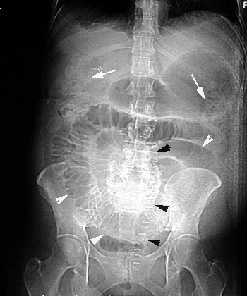
Plain film in a case with Small bowel obstruction
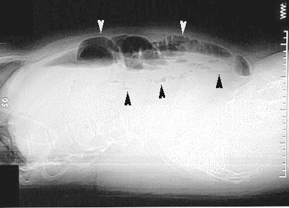
  • Multiple dilated small bowel loops are seen (white arrowheads).
  • There is fecal material in the right and left colon
    (arrows).
  • Air is seen in the rectum.
  • The surgical staples indicate recent abdominal surgery. (black arrowheads).


Small bowel obstruction

Cross lateral view shows multiple dilated fluid filled loops of bowel with air fluid levels.

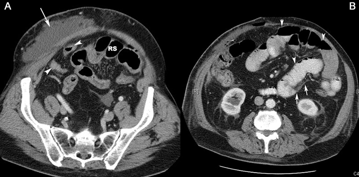
CT scan of another patient showing findings of small bowel obstruction:
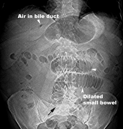
  • A: Post op changes in abdominal wall. Arrowheads: Normal size of distal small bowel and Recto sigmoid (RS)
  • B: Arrowheads: Dilated small bowel loops. Arrow: Area of obstruction

Gall stone Ileus

  • Air in Bile duct
  • Dilated small bowel
  • Gall stone is not seen in this film and may overlie the sacrum where the dilated bowel seems to end. [Black arrow ]


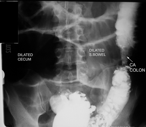
Lower GI in a patient with Large bowel Obstruction

  • Dilated bowel loops proximal to the obstruction.
  • Arrow points to the etiology of obstruction.