Kidneys
Location:
Located in the posterior abdomen, paravertebraly in the retro peritoneum
They are situated at the level of the costo-veretebral angle
The right kidney is 2-8 cm lower than the left Kidney, because of the large liver which sits superior to it.
Shape:
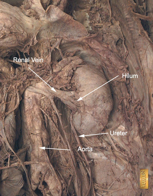
Hilum
Hilus leads into the renal sinus which contains renal artery, vein and renal pelvis.
Renal vein anteriorly, Renal artery in middle and Renal pelvis posteriorly
Size:
Measures 9-11 cm in length x 6 cm in Width x 3 cm in AP
US is the best modality to obtain the size of kidneys
Contour
Smooth
Axis
Long axis - directed outwards and laterally
Tilted, superior pole closer to mid line
Relationship:
Anteriorly the right kidney is related to the Liver, duodenum and hepatic flexure of ascending colon
Anteriorly the left kidney is related to Stomach, Jejunum, Pancreas, Spleen and descending Colon
Adrenal gland is superior and anterior to the Kidneys
The kidney is surrounded by a distinct layer of fascia (Perirenal fascia) that separates the fat surrounding the kidney into the perinephric and paranephric fat. Kidneys lie in a fatty cushion of perinephric fat
Right Kidney is related to the 12th rib posteriorly resting on diaphragm
The left kidney rises to as high as the 11th thoracic rib posteriorly, extending from T11-L3, resting on diaphragm
Thin capsule
Renal cortex
Renal cortex consists of glomeruli and renal tubules
Normal thickness is 2.5 cms
Consists of multiple renal pyramids which have their base to the periphery and their conical end directed towards the renal hilus.
Their tips are called papillae.
Each minor calyx receives 1-3 papillae, through which the collecting tubules empty.
Columns
Embryology:
The kidneys develop in the pelvis and ascend up.
If arrested, they can be seen in various portion of their route.
They may fuse to form a horseshoe kidney.
Applied Anatomy