![]()
There are several common situations that potentially cause a problem if not anticipated. Proper preparation through education and drills should prevent delays in patient care. The most common of these special situations is as follows:
The patient is contaminated, not yet stable, and needs radiographs of various regions to help make decisions. Cervical spine x-rays may be needed to exclude the presence of fracture. A chest x-ray may be needed to determine the placement of a central line or evaluate for fractured ribs or widened mediastinum. Since the patient is contaminated and unstable, it is unwise to move the patient. A portable x-ray machine can be used to obtain many of the required diagnostic images.
The portable x-ray machine can be rolled into the buffer zone and the patient brought close to the doorway. The film cassette is covered with a plastic garbage bag or pillowcase and then positioned as usual. The portable x-ray head is placed in the correct position for the radiograph and the image obtained.
When the cassette is brought out of the room, the buffer zone nurse can grab the clean cassette from inside the covering. This virtually assures that the cassette is not contaminated. Nevertheless, it should be checked by a radiation technologist before sending it to be developed. This procedure can be repeated for all necessary x-rays. When complete, the wheels and the mobile head of the x-ray machine should be checked for the presence of contamination. As long as the machine remained in the buffer zone, it should still be uncontaminated.
Alternatively, a white sheet of Herculite can be rolled into the room as a pathway for the x-ray machine. The staff in the REA should not walk over or contaminate the clean pathway. The x-ray machine can then be brought into the room on the white sheet and the radiographs taken. When complete, the machine can be rolled out on the pathway. Again the wheels should be checked for areas of contamination.
If the REA is large enough, and a large number of radiographs are anticipated, the x-ray machine can be rolled into the REA without any of the above precautions and kept there for the duration of patient care. When the patient is stabilized, decontaminated and moved to a definitive care center, the machine can be checked for areas of contamination and cleared for general use by a radiation technologist.
Should an x-ray cassette become contaminated regardless of the protection by a plastic bag or pillowcase, it should be handed back into the room and the affected area washed to remove the contamination. This is generally easily removed by the use of soap and water. Once it is decontaminated, it can be sent for processing.
These studies present more of a problem. The size of these scanners prohibits them from coming to the patient. In this case, the patient must be prepared for transport to a scanner.
First, the patient should be stabilized as usual and the radiology department notified that a patient will be arriving for a particular type of scan. Often, there will be some delay until the scanner is available. During this time, further stabilization efforts, specimen collection, and decontamination can begin. If the scanner becomes available while the patient is still contaminated, the patient should be cocooned in sheets or the contaminated areas wrapped up with bandages. In this way, the patient will not be able to contaminate other equipment or personnel.
The patient can then be brought out of the REA using a clean team transfer technique as described below. The CT or MRI scan can be performed as usual, and when finished, can be brought back into the REA for further management and decontamination.
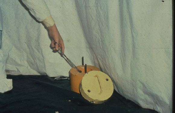
Depending on the type of accident and the materials involved, it is possible that a piece of radioactive material with higher activity may be present on the patient. This does not usually present a significant safety problem for the staff involved. The location of the particle can easily identified by radiation technologists. The important point to remember is not to pick it up with fingers. The inverse square law applies in reverse as well. The closer to the source, the greater the exposure. Once identified, the hot particle can be removed using either a forceps or by using a piece of tape. Once identified and removed from the patient, it can be placed within a lead pig (a lead container with thick walls) to prevent further exposure to the staff. Below is a photograph of a lead pig.

It is possible that the nature of the wounds may require urgent surgical intervention. Although this presents a potential contamination problem, it is simply addressed. The operating suites usually require some time to prepare for surgery. During this time, efforts toward stabilization should be performed. If time permits, sample collection and a decontamination effort can also begin. Full decontamination efforts however will probably not be complete at the time of surgery. Areas that cannot be decontaminated can be bandaged and labeled as contaminated.
A clean team transfer can be performed to transfer the contaminated patient to a new cart and then transported to the surgical suite. Once the patient arrives in the operating room, the suite becomes the new REA. Nothing should leave the new REA without being checked for contamination. Once surgery is completed, decontamination efforts can beresumed. The patient can then be transferred to a definitive care area.