Abnormal Finding
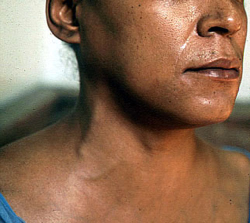
Neck vein distension at 45 o inclination is abnormal and is indicative of increased central venous pressure. Describe the level to where the pulsations are seen in relationship to the angle of Louis. Note the effect of inspiration. Apply gentle pressure to right upper quadrant and note its effect on neck veins (hepatojugular reflux). If neck vein distension is present identify a, c and v waves and describe their amplitude.
- Distended pulsatile neck veins ( CHF, Tricuspid insufficiency)
- Hepatojugular reflux: Right ventricular non-compliance to increased filling
- Distended non- pulsatile neck veins: ( SVC syndrome , cardiac tamponade, Constrictive pericarditis). These patients usually have prominent descents.
- Quick Y descent and X descent: (Constrictive pericarditis)
- Distended veins during expiration only: (COPD, Asthma)
- Prominent "a" wave: "a" waves are due to atrial contraction and when abnormally prominent indicate atrial contraction into a noncompliant right ventricle or through a stenotic or closed tricuspid valve. In complete heart block and with premature ventricular contraction there is loss of a-v synchrony. When the atrial and ventricular contractions coincide a prominent wave is seen. This is called cannon a-wave. A noncompliant right ventricle can be hypertrophied (secondary to pulmonary hypertension) or "stiff" due to scar (ischemia/infarct) or infiltrative disease (amyloid).
- JVP which increase with inspiration indicate restricted filling of the right sided chambers (Kussmaul's signs).
- Absent "a" waves: (Atrial fibrillation).
- "v" waves are most commonly due to an insufficient tricuspid valve with the ventricular systolic pressure reflected in the atrium during atrial filling (diastole).
- Prominent "v" wave: (Tricuspid regurgitation).
- Cannon wave: (Heart block, Premature ventricular contraction).