This is the current version of Pulmonary artery and vein 06/29/00
Read first the text book: Computed Tomography and Magnetic resonance of the Thorax by Nadich et all
Then go through this exercise to assess your comprehension
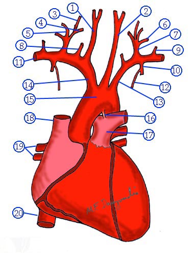
Pulmonary artery
Q: Which component is the reference landmark and why?
Airways are the reference landmarks Air in lumen serves as a contrast agent for easy recognition
Constant relationship of pulmonary artery and vein to airways
Q: What is the extent, length, orientation, diameter and branches of main pulmonary artery?
Curves upwards from right ventricle
Forms a segment of left Heart border
Bifurcates at the level of Carina slightly left of midline
Q: What is the extent, length, orientation and diameter of right branch of pulmonary artery?
Is horizontal, Horizontal
Divides into two major branches in mediastinum
Lies posterior to Aorta and the superior vena cava
Anterior to right main bronchus
Divides into Truncus anterior and Interlobar artery
Truncus anterior supplies RUL
Curves anteriorly and superiorly over upper lobe bronchus
Divides into three branches corresponding to bronchial segments
Interlobar artery
Is larger and passes in front of and along lateral side of intermediate bronchus
Transverse diameter measures 15-16 mm during inspiration
Enters into oblique fissure
Supplies RML and segments of Lower lobe
Q: What is the extent, length, orientation and diameter of left branch of pulmonary artery?
Curves upwards and backwards towards hilum
Passes over anterior and lateral to left main bronchus
Slightly higher and posterior than right
Left hilum higher than right in 97% of normals
Divides into two branches in hilum
Upper division supplies segments (apical-posterior and anterior) of LUL
Interlobar artery
Curves sharply over the top of LUL and descends along lateral aspect of LLL bronchus
Enters Oblique fissure
Lingular branch arises from anterior aspect
Superior segmental artery from posterior aspect
Supplies branches to LLL basal segments
Pulmonary Veins
Q: Describe the venous drainage of lungs
Two on either side
Superior and inferior
Enter the mediastinum slightly below the pulmonary arteries and anterior to them
Right superior vein drains RUL and RML
Right inferior vein drains RLL
Left superior vein drains LUL and Lingula
Left inferior vein drains LLL
Q: What are the distinguishing characteristics between veins and arteries
Course of veins is remote from artery
Veins begin in lung periphery
Arteries are in center of lobules
Veins and arteries are separated by lungs
Veins run lateral or inferior to respective arteries
Horizontal topography of veins while arteries have a vertical course especially in lower lobes
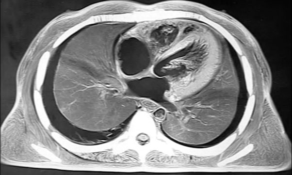
CT recognition
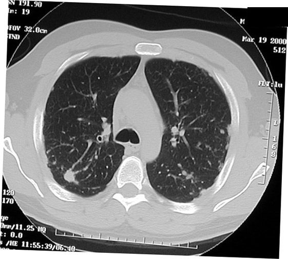
Q: Click to look at the section at the level of Carina. Identify the labeled structures.
Circular lucency( Apical segmental bronchus of RUL) in association with artery and vein
RUL pulmonary artery. Artery lies just medial to bronchus
Right superior pulmonary vein. Vein just lateral to bronchus
Q: You are looking at section at the level of RUL bronchus (at or just below carina). Identify the labeled structures.
RUL bronchus originates more cephalad than LUL bronchus
Above the right main pulmonary artery
Posterior wall is in contact with either posterior segment of RUL or superior segment of RLL
Truncus anterior first major branch of right pulmonary artery lies anterior to RUL bronchus
Branch of right superior pulmonary vein lies at the angle between anterior and posterior segmental bronchi
Anterior and medial to truncus anterior lies RUL vein (the apical-anterior vein)
Q: You are looking at section at the level of bronchus intermedius. Identify the labeled structures.
Lies directly behind right main pulmonary artery
Medial to right intralobar pulmonary artery
Posterior wall in contact with superior segment of RLL
Azygoesophageal recess is along posteromedially separated by lung tissue
Inferior to bronchus right superior pulmonary vein lies alongside the lateral border of right interlobar pulmonary artery
Multiple veins can be identified in this location
Right main pulmonary artery at the lateral border of intermedius bronchus turns inferiorly and becomes interlobar pulmonary artery.
At this position the artery may have a triangular configuration
Q: You are looking at section at the level of RML bronchus Identify the labeled structures.
Interlobar pulmonary artery is vertical and lies immediately lateral to RML and RLL bronchi
Artery has an oval configuration at this point
Right superior pulmonary vein lies medial to RML bronchus and can be seen entering the upper portion of left atrium
Q: You are looking at section at the levell of RLL basilar segments Identify the labeled structures.
Lower lobe lies medial and anterior to lower lobe pulmonary artery
Appears suspended by superior portion of the inferior pulmonary ligament
Medial basilar segment arises first, anterior to inferior pulmonary vein
Branches of right interlobar pulmonary artery are round and lay posterolateral to the proximal portions of basilar segments
Inferior pulmonary veins are oriented horizontally and can be traced to right inferior pulmonary vein and subsequently into the lower portion of left atrium.
Q: You are looking at section at the level of lower trachea and carina. Identify the labeled structures.
Apical-posterior segmental bronchus is separated from the left main bronchus by the left main pulmonary artery, which courses over LUL bronchus at this level
LUL pulmonary artery cab be traced to its origin from the left main pulmonary artery and lies posterolateral to the left superior pulmonary vein
Superior pulmonary vein tends to be located anteriorly
Q: Left upper lobe
Forms a sling over which the main pulmonary artery passes
Q: You are looking at section at the level of upper portion of LUL Identify the labeled structures.
Posterior wall is smooth and slightly concave caused by the left pulmonary artery
Superior segment of Lower lobe abuts the posterior portion
Posterior wall of LUL bronchus is slightly convex because it is indented superiorly and posteriorly by the left main pulmonary artery
Posterior to LUL bronchus the left pulmonary artery continues as interlobar pulmonary artery
Appears triangular at this level because of the change in course
Left superior pulmonary vein lies in front of LUL bronchus and has a horizontal course
Distinct branching can be identified peripherally
Q: You are looking at section at the level of lower portion of LUL Identify the labeled structures.
Left interlobar artery has a vertical course and lies lateral to the spur
Lateral to the origin of the lingular bronchus the pulmonary artery to lingula can be identified
Directly anterior to LUL bronchus is the inferior portion of the left superior pulmonary vein
Pulmonary parenchyma can be seen between pulmonary artery and descending aorta (retro bronchial stripe)
Q: You are looking at section at the level of LLL basilar segments Identify the labeled structures.
Branches of pulmonary artery to the lower lobe lie lateral and posterior to the basilar bronchi
Left inferior pulmonary vein is horizontal and cab traced to left atrium
Left inferior pulmonary vein course along the lateral border of descending aorta
Read first the text book "Synopsis of Diseases of the chest by Pare and Fraser" by W.B.Saunders company
Roentgenology of the vascular system.
The session is over