|
UNDER CONSTRUCTION This has two modules
|
|
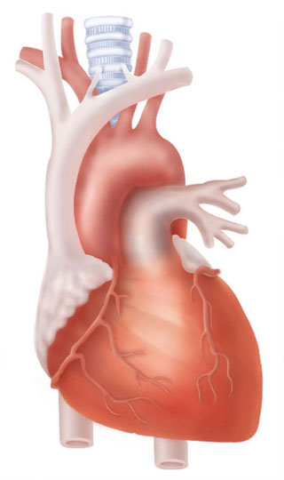
| Heart | |
| Right atrium | |
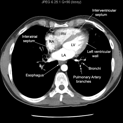
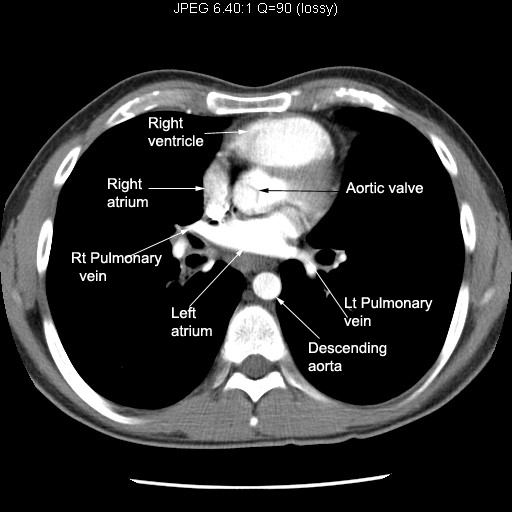
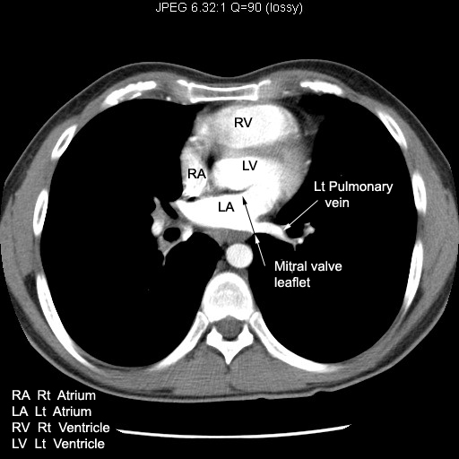
| Location | Identify right atrium |
| Receives | CT: Follow SVC entering right atrium. |
| Empties into | CT: Right atrium emptying into right ventricle. |
| Tricuspid Valve | Tricuspid valve permits blood flow into right ventricle |
| Relationship | CT: Note the relationship of right atrium to other chambers of heart. |
| Heart Border | CT: Note which heart border, the right atrium forms |
| Appearance in | Angiogram |
| CXR: Identify the location of right atrium | |
| Ultrasound | |
| MR: | |
| Left atrium | |
| Location | |
| CT: Pulmonary veins entering left atrium | |
| Empties | CT: Left atrium emptying into left ventricle |
| Mitral valve | CT: Note mitral valve leaflet. Note its location. |
| Relationship | CT: Relationship of LA to Esophagus |
| Barium Swallow: Note the indentation of Esophagus by left atrium | |
| CXR: Left main stem bronchus along the superior margin of left atrium. | |
| Heart Border | CXR: Note left atrial appendage forming a portion of left heart margin. |
| Left atrium forms the posterior margin of heart. | |
| Appearance in | Angiogram |
| Ultrasound | |
| MR: | |
| Right ventricle | |
| Location | Locate right ventricle |
| Location | Locate right ventricle |
| Receives | Blood from right atrium |
| Tricuspid valve separates right atrium from right ventricle | |
| Empties | CT:Right atrium emptying into right ventricle. |
| Pulmonary artery originates from right ventricle | |
| Pulmonary valve separates right ventricle from pulmonary artery | |
| Relationship |
CT: Note the relationship of right ventricle to Sternum. |
| Relationship | CT:Note the relationship to other chambers of heart |
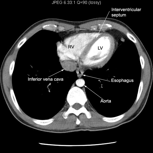
| Septum | CT:Note the inter ventricular septum |
| Appearance in | Angiogram |
| CXR: Locate right ventricle | |
| Ultrasound | |
| MR: | |
| Left ventricle | |
| Location | |
| Receives | Left atrium empties into left ventricle |
| Mitral valve separates left atrium from left ventricle. | |
| Chorda tendinae and papillary muscle anchor mitral valve to left ventricle | |
| Empties | Aorta originates from left ventricle |
| Aortic valve separates left ventricle from Aorta | |
| Wall thickness |
CT: Note left ventricular wall thickness and Inter ventricular septum compared to right ventricle |
| Heart margin | CT: Forms apex of heart and part of left heart border |
| Relationship | |
| Appearance in | Angiogram |
| CXR: Locate left ventricle | |
| Ultrasound | |
| MR: | |
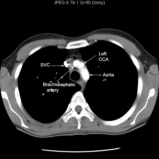
| Aorta | |
| Thoracic Aorta | |
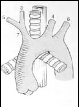
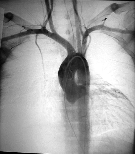
| Drawing | Follow Aorta from Aortic valve, ascending, arch and descending portions of Aorta. |
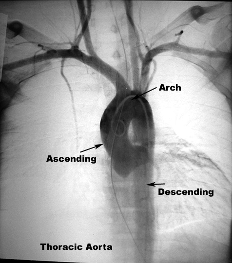
| Angiogram | Follow Aorta from Aortic valve, ascending, arch and descending portions of Aorta |
| CXR | Follow various portions of Aorta |
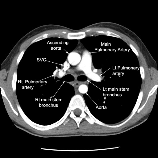
| Ascending aorta | |
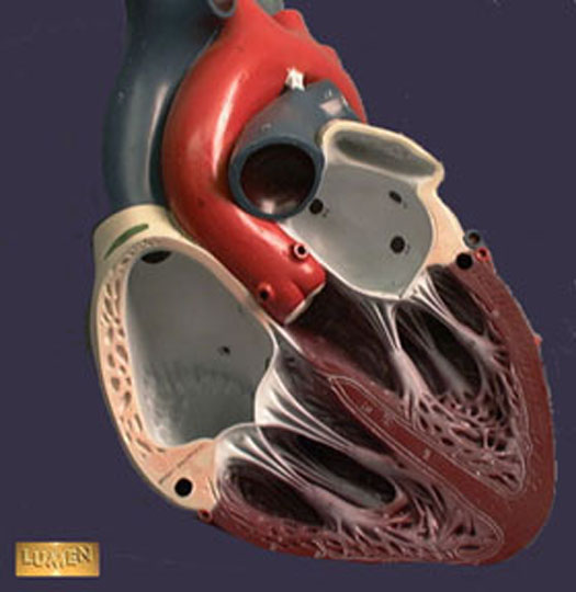
| Model | Note the origin of Aorta from Left ventricle and its relationships. |
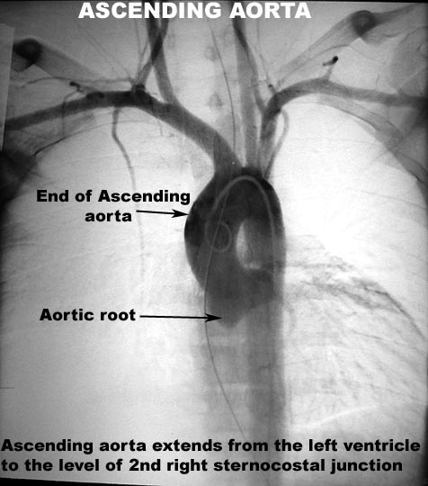
| Course | Angiogram: Ascending Aorta Start and end |
| Aortic valve | CT: Aortic valve |
| Branches | Angiogram: Branches of Ascending Aorta |
| Relationship | CT: Main pulmonary artery is to left of aortic root. |
| Appearance in | CXR |
| MR | |
| Angiogram | |
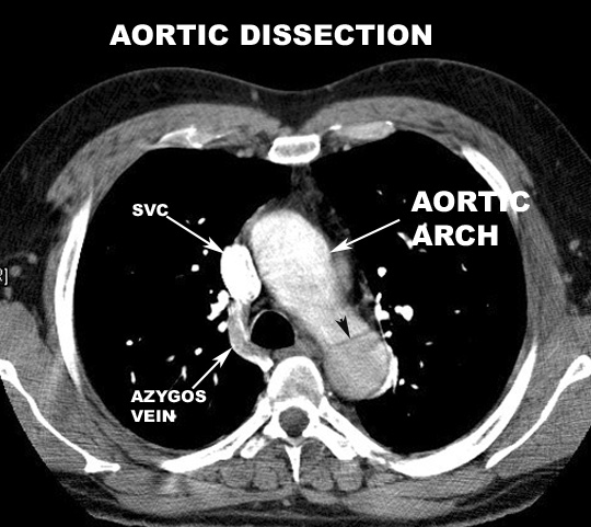
| Arch of aorta | |
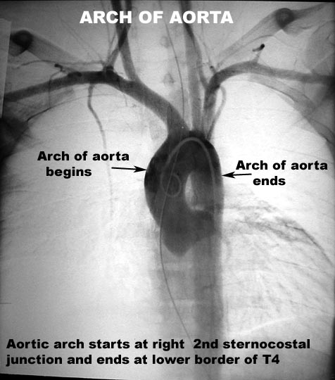
| Course | Angiogram: Start and end of arch of aorta. |
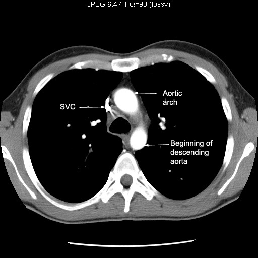
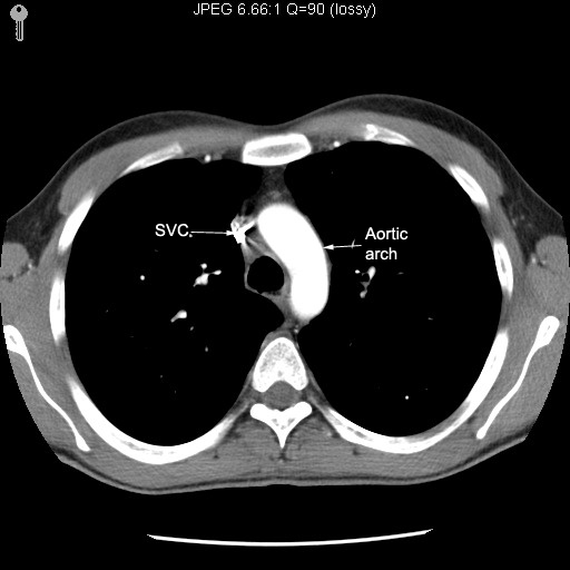
| Origin | CT: Start of aortic arch |
| Course | CT: Aorta arching across mediastinum |
| Branches | Angiogram: Branches of arch of aorta |
| Branches | Major branches of Thoracic Aorta. |
| Branches | CT: Major branches on the surface of aortic arch |
| Relationship | |
| Appearance in | CXR |
| MR | |
| Angiogram | |
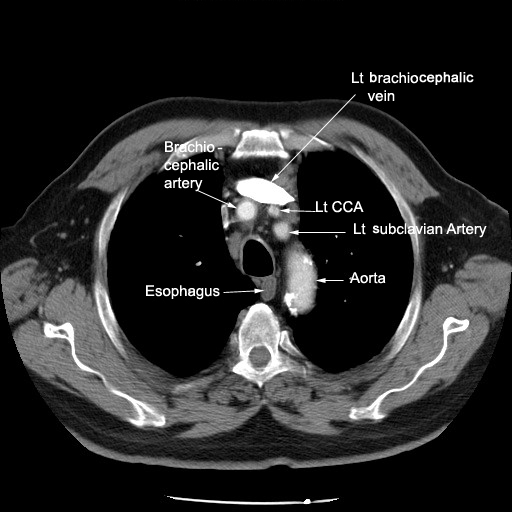
| Descending aorta | |
| Course | Angiogram: Aorta and branches |
| Relationship | CT: Relation to Esophagus, left pleura and Vertebra |
| Relationship | CT: Relation to Esophagus, left pleura and Vertebra |
| Relationship | CT: Relation to Esophagus, left pleura and Vertebra |
| Appearance in | CXR |
| CXR: Locate various portions of Aorta | |
| MR | |
| Angiogram | |
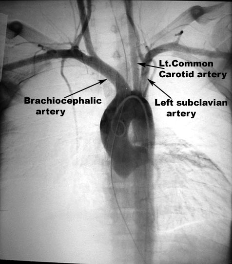
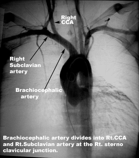
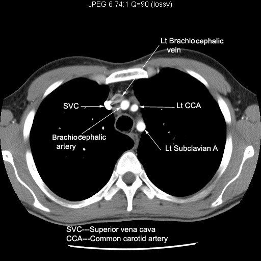
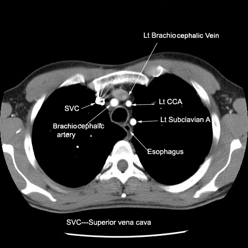
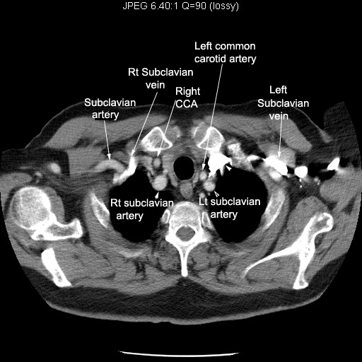
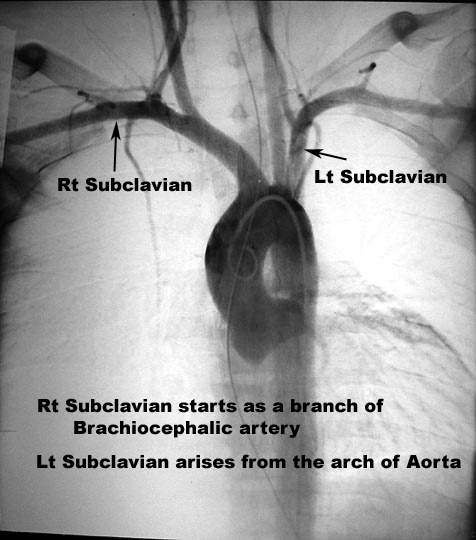
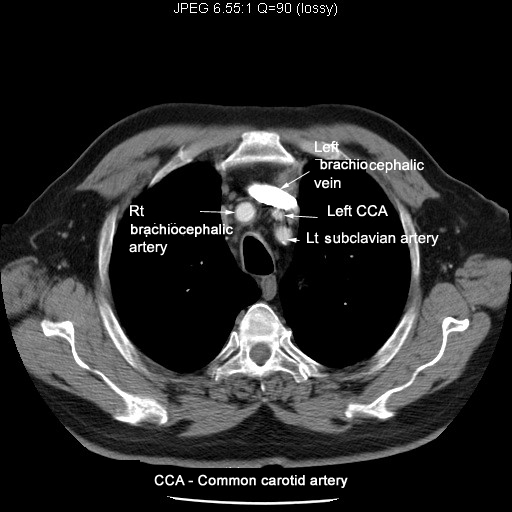
| Brachio cephalic artery | |
| Course | Angiogram: Aorta and branches |
| Dissection | |
| Origin | CT: Starting from the arch of aorta |
| Relationship | CT:Relation to brachiocephalic vein |
| Relationship | CT:Relation to brachiocephalic vein |
| Branches | CT:Divides into right common carotid and right subclavian arteries |
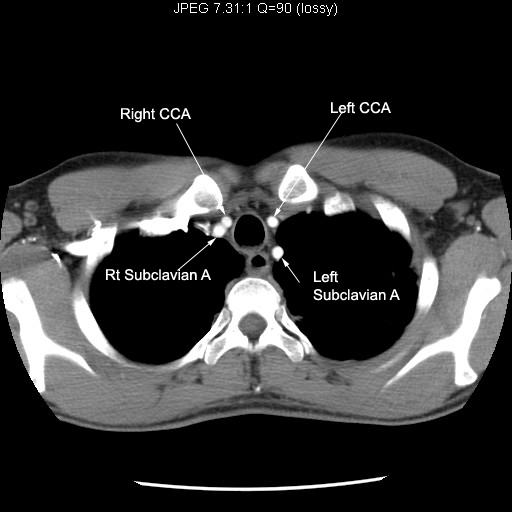
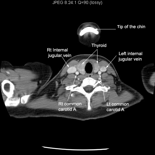
| Left Common carotid artery | |
| Course | Angiogram: Aorta and branches |
| Origin | CT:Starting from the arch of aorta |
| Origin | CT:Middle branch of the arch of aorta |
| Origin | CT:Middle branch of the arch of aorta |
| Relationship | CT:Courses towards neck |
| Relationship | CT:Courses into neck |
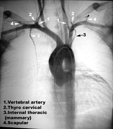
| Branches | |
| Course | Angiogram: Origin of subclavian |
| Branches | Angiogram: Branches of subclavian artery |
| Origin | CT: Lat branch off of arch of aorta |
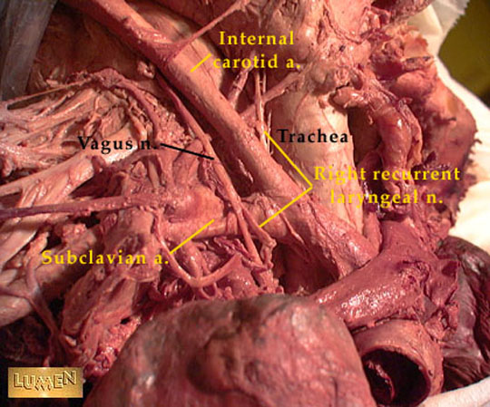
| Relationship | CT:Relation to trachea and esophagus |
| Relationship | CT:Relation to trachea and esophagus |
| Relationship | CT:Coursing into neck |
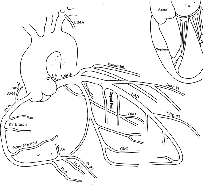
| Coronary artery | |
| Branches | Drawing: Coronary artery and its branches |
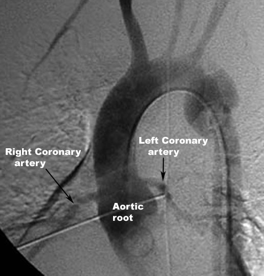
| Origin | Angiogram: Branch of Ascending Aorta |
| Angiogram | Left Coronary artery |
| Angiogram | Right Coronary artery |
| Angiogram | Branches of LCA |
| Angiogram | Branches of RCA |
| Superior Vena cava | |
| Location | SVC entering right atrium |
| Origin | CT: Left joining right brachiocephalic vein to form SVC |
| Origin | CT:Left joining right brachiocephalic vein to form SVC |
| Relationship | CT:Relationship to Aorta and right pulmonary artery |
| Ends | CT:SVC drained into right atrium |
| Tributaries | CT:Azygous vein joining SVC |
| Appearance in | |
| Angiogram | |
| CXR: Locate SVC | |
| MR | |
| Nuclear Medicine | |
| Brachiocephalic veins | |
| Formation | CT: Internal jugular vein joins subclavian vein to form brachiocephalic vein |
| Formation | CT: Internal jugular vein joins subclavian vein to form brachiocephalic vein |
| Formation | CT: Left joins right brachiocephalic vein to form SVC |
| Course | CT: Left brachiocephalic crosses mediastinum towards right brachiocephalic vein |
| Course | CT: Right brachiocephalic descends vertically |
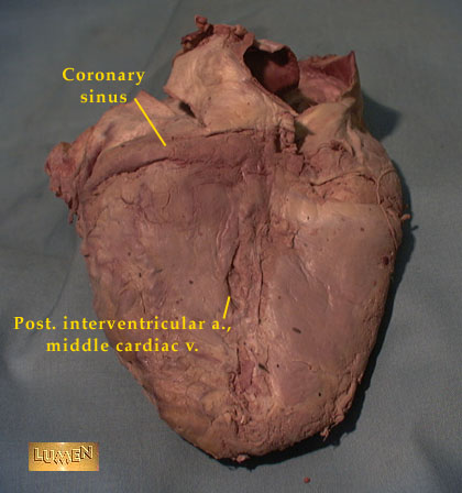
| Location | Coronary sinus |
| Location | Coronary sinus in the atrioventricular groove and entering right atrium |
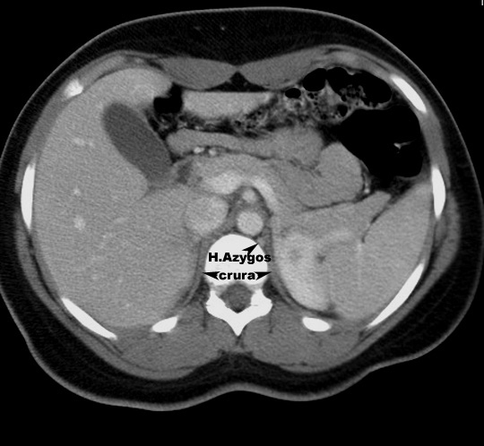
| Location | CT: Locate Azygous vein and identify the nearby structures |
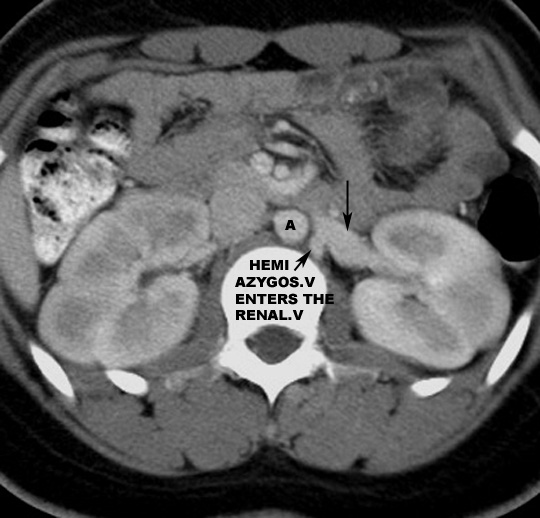
| Location | CT:Locate Azygous and Hemiazygous veins. |
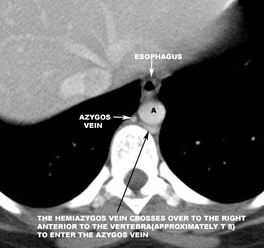
| Relationship | CT:Azygous vein relation to Aorta, Vertebra and Esophagus |
| CT:Note the relationship of Hemiazygous vein relation to Aorta, Vertebra and diaphragmatic crus | |
| CT:Hemiazygous connecting to left renal vein in this case. | |
| CT:Hemiazygous crosses over to join Azygous vein | |
| Course | CT:Azygous joining SVC |
| Appearance in | |
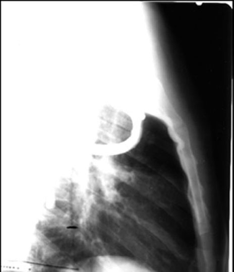
| Venogram Patient with mediastinal fibrosis and obstructed SVC, showing large azygous vein entering SVC | |
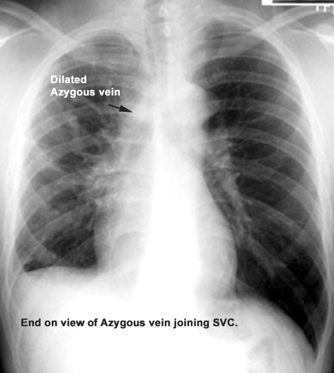
| CXR Patient with mediastinal fibrosis and obstructed SVC, showing large azygous vein entering SVC | |
| Inferior vena cava /Thoracic Portion | |
| Location | |
| Course | |
| Appearance in | CXR: Locate IVC |
| Lateral CXR: Locate IVC | |
| CT: IVC entering RA | |
| Pulmonary artery | |
| Location | Locate main pulmonary artery. |
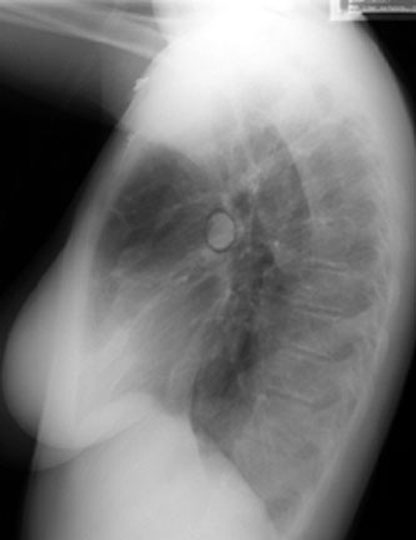
| Course | CXR Pneumo-mediastinum outlining right pulmonary artery. End on view. |
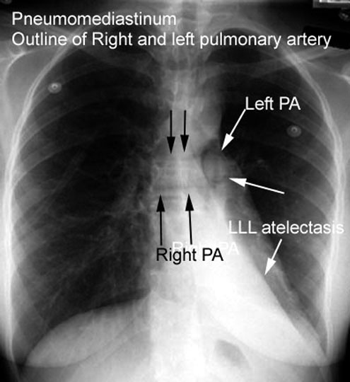
| Course | CXR: Pneumo-mediastinum outlining left and right pulmonary arteries. |
| Relationship | CT: Maim pulmonary artery to the left of Aorta |
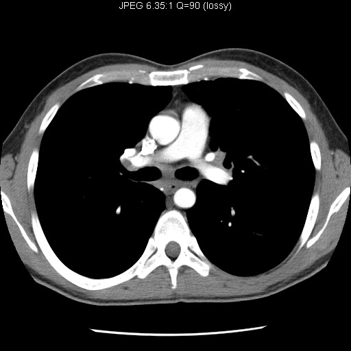
| Branches | CT: Divides into left and right pulmonary arteries |
| Branches |
CT: Pulmonary artery branches in the lungs. |
| Appearance in | |
| Angiogram: Main Pulmonary artery | |
| Angiogram: Right Pulmonary artery | |
| Angiogram: Left Pulmonary artery | |
| Angiogram: Lateral view | |
| CXR: Locate pulmonary arteries | |
| Lateral: Locate left and right pulmonary arteries | |
| Pulmonary veins | |
| Course | CT: Pulmonary veins entering left atrium. |
| Course |
CT: Left Pulmonary veins entering left atrium. |
| Appearance in | |
| CXR: Locate pulmonary veins | |