Hematuria / Renal Stone
- Trauma
- Renal calculi
- Renal cancer
- Bladder stone or cancer
- Acute glomerulonephritis
Urine analysis is diagnostic showing red cells and red cell casts in acute glomerulonephritis
What is the common cause for painful hematuria?
- Calculi in GU tract
What is the common cause for painless hematuria?
- Renal cancer
What are useful imaging modalities to investigate renal stones?
- KUB plain film
- Non contrast CT abdomen
- Ultrasound
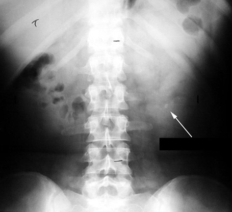
KUB Plain film
Kidney Stone
Abdominal radiograph of the kidneys, ureter and bladder (KUB)- Allows for the evaluation of urinary tract stones.
- Ninety percent or more of urinary-tract stones are radio-opaque.
- The degree of opacity varies depending upon the composition of stone (calcium content of the stone).
- Uric acid stones (8%) are radiolucent.
- Visibility of stones depends not only on the degree of opacity, but also on their sizes and positions relative to other abdominal structures.
- An opaque stone needs to be approximately 2mm in its largest diameter to be visible on an abdominal film.
- Poor sensitivity for ureteral stones.
Non-contrast helical computed tomography of the abdomen (NCHCT)
- Greater sensitivity than KUB for ureteral stones
- Most sensitive and specific (95-97% and 96-97% respectively)
- Because abdomen and pelvis are scanned in one to two breaths, virtually eliminates the problem of respiratory motion
- Can be performed quickly, eliminating potential delays
- Safe, because eliminates the risk of contrast, which includes allergic reactions and toxicity
- Detects non-urologic pathology - including appendicitis or ovarian cysts
- While more expensive than a KUB, comparable to a excretory urography and ultrasound
- Able to detect all stones regardless of composition, except those associated with indinavir therapy for HIV infections
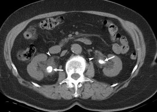
Bilateral Renal Pelvis Calculi
Arrowheads: There is soft tissue infiltration secondary to extravasations of urine around both renal pelvis.
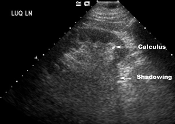
Ultrasound
- Preferred procedure in pregnant women and in patients allergic to IV contrast.
- Not dependent on the composition of stones and detects uric-acid stones as well as calcium stones.
- Stones are seen as highly echogenic foci and often produce distal acoustic shadowing
- Not always possible to distinguish small stones from arterial calcifications, pericalceal fat, or crystal-laden calceal sub mucosal plaques.
- Detects hydronephrosis
- Generally good sensitivity .
- Ureteral stones are difficult because of overlying gas
Kidney Stone
US: Lower pole calculus.
Sensitivity, specificity of procedures.
|
PROCEDURE |
SENSITIVITY |
SPECIFICITY |
|
KUB |
45-70% |
77% |
|
CT |
95-97% |
96% |
|
Ultrasound |
32-70% |
70-97% |
Utility of imaging procedures
Indicate when you would select each procedure.
Controversy exists over the proper procedure sequence. With the addition of non-contrast helical CTs, some advocate CT as the first-line procedure. However, most physicians continue to utilize plain radiographs of the abdomen (KUB) as the initial screening imaging procedure.