|
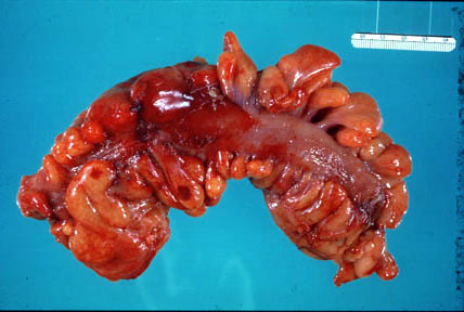
Acute diverticulitis occurs when a section of colon reveals acute inflammation (hyperemia, swelling) of the serosa and pericolic fat. |
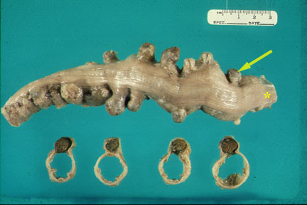 |
A section of colon reveals numerous diverticula which protrude from the edge of the taenia coli (*). The colon is cut in cross section revealing the diverticuli (contain feces) and the empty colonic lumen. |
 |
Diverticula |
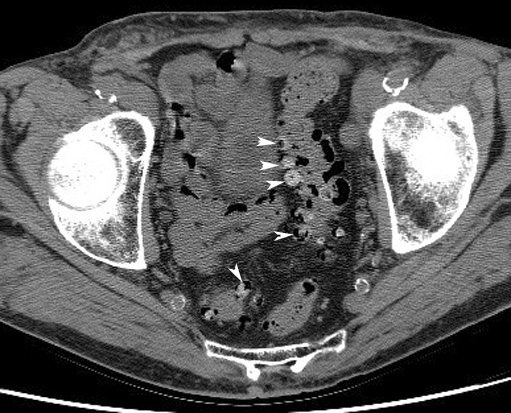 |
DiverticulaCT: Arrowheads point to multiple diverticula arising from the rectosigmoid. |
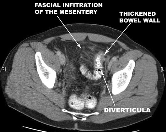 |
Diverticulitis |
|
|
Pneumoperitoneum
|
 |
Diverticulitis with AbscessFindings:
|
