|
There are two modules in this section:
UNDER CONSTRUCTION
|
|
| This exercise is intended for students studying Anatomy. Click the links in the left-hand column to view the unlabled images, and try to answer the questions using this image. When you have your answers, click the image to reveal the labels and check your answers. | |
| CT Densities | |
| CT | Identify the densities in CT. |
| CT | Identify the densities in CT. |
| Location | |
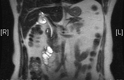
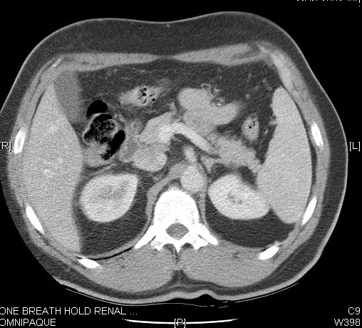
| CT | Locate the gall bladder. |
| CT | Locate the gall bladder. |
| Parts | |
| Cholecystogram | Identify the parts of the gall bladder. |
| Relationship | |
| CT | Note the relationship of the gall bladder to other structures. |
| Appearance | |
| US | Locate the gall bladder in the ultrasound. |
| US | Locate the gall bladder in the ultrasound. |
| MRCP | Identify the components of the biliary drainage system. |
| Operative Cholecystogram | Identify the components of the biliary drainage system. |
| Hepatic Duct | |
| Cystic Duct | |
| Pancreatic Duct | |
| CT | Locate the pancreatic duct. |
| CT | Locate the pancreatic duct. |
| Common Bile Duct | |
| CT | Locate the common bile duct. |
| CT | Note the location of CBD in relation to the duodenum. |
| MR | GB and CBD |
| Ampulla of Vater | |
| Location | |
| CT | Locate and identify the pancreatic body and tail. |
| Parts | |
| CT | Identify the uncinate process. |
| CT | Identify the uncinate process of the pancreas. |
| CT | Note the tail of the pancreas in the hilum of the spleen. |
| CT | Note the tail of the pancreas in the hilum of the spleen. |
| Pancreatic Duct | |
| CT | Locate the pancreatic duct. |
| Relationship | |
| CT | Note the location of the pancreas in relations to the duodenum . |
| CT | Note the relationship of the pancreas to surrounding structures. |
| CT | Identify the vessels around pancreas. |
| CT | Identify the vessels around pancreas. |
| CT | What is the relationship of the pancreas to the splenic vein? |
| CT | Note the relationship of the pancreas to surrounding structures. |
| CT | Identify the vessels around the pancreas. |
| CT | What is the relationship of the pancreas to SMA and SMV? |
| US | Note the relationship of splenic vein to the pancreas. |
| Appearance | |
| Color Doppler | Note the pancreas with vascular landmarks. |
| CT | Locate the lesser sac. |
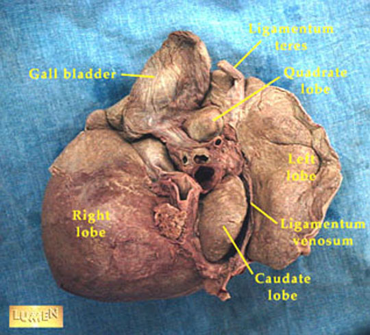
| Dissection | View the lobes of the liver. |
| Location | |
| CT | Locate Porta Hepatis. |
| Lobes / Parts | |
| CT | Identify the right, left and caudate lobes of the liver. |
| CT | Identify the right and left lobes of the liver. |
| CT | Identify the caudate lobe. |
| CT | Identify the fissure for ligamentum teres and venosum. |
| CT | Segments of liver |
| CT | Hepatic veins as a marker of lobes of liver |
| CT | Identify fissures |
| CT | Identify the segments of the left lobe of the liver. |
| Relationship | |
| CT | Identify the structures around the liver. |
| CT | Identify the vessels in the Porta Hepatis. |
| Appearance | |
| CT | Note anything unusual about this liver. |
| Location | |
| CT | Locate the spleen. |
| CT | Locate the spleen and surrounding structures. |
| CT | Locate the spleen and adrenal glands. |
| Parts | |
| Relationship | |
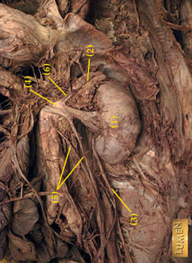
| CT | Identify the structures in the hilus of the spleen. |
| CT | Identify the structures in the hilus of the spleen. |
| CT | Identify the splenic vein. |
| CT | Identify the round structure between the spleen and the tail of the pancreas. |
| Appearance | |
| US | Note the spleen in this ultrasound. |
| Adrenals | |
| Anatomy | Note the location of the adrenals. |
| Location | |
| CT | Locate the adrenal glands in relation to the kidneys. |
| CT | Locate the adrenal glands. |
| CT | Locate the adrenal glands. |
| CT | Note the location of the adrenal glands. |
| CT | Locate the right and left adrenals. |
| CT | Locate the right and left adrenals. |
| CT | Locate the right and left adrenals. |
| CT | Locate the adrenal glands. |
| Right Adrenal | |
| CT | Locate the right adrenal gland. |
| CT | Note the shape of the right adrenal gland. |
| Left Adrenal | |
| CT | Note the shape of the left adrenal. |
| Relationship | |
| CT | Identify the structures around the adrenal gland. |
| CT | Identify the structures around the adrenal gland. |
| CT | What is the relationship of the adrenals to the kidney? |
| Location | |
| Openings | |
| Left hemidiaphragm | |
| CT | Relationship of left crus of diaphragm |
| Right Hemidiaphragm | |
| CT | Relationship of right crus of diaphragm |
| Relationship | |
| CT | Identify the structures around the diaphragmatic crura |
| CT | Identify the structures around the diaphragmatic crura |
|
Abdominal Muscles |
|
| CT | Identify the muscles. |
| CT | Identify Iliacus. |
| CT | Identify the muscles. |
| CT | Identify the muscles. |
| CT | Identify obturator internus. |
| CT | Identify the muscles. |
| CT | Identify Levator ani. |
| CT | Identify the muscles. |
|
Pelvis |
|
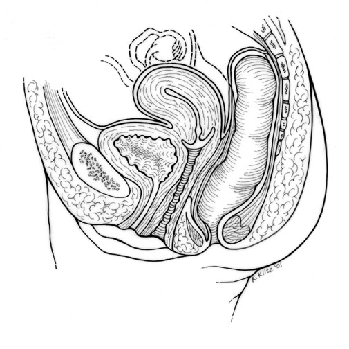
| Drawing | Female |
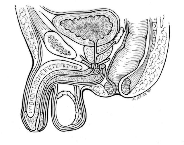
| Drawing | Male |
| Uterus | |
| CT | Identify the Uterus and the structures around |
| CT | Identify the Uterus, Ovaries and the structures around |
| US | Myoemetrium and Endometrial stripe |
| US | Uterus and Ovary |
| US | |
| US | |
| Hysterosalpingogram | Uterus and Fallopian tubes |
| Hysterosalpingogram | Uterus and Fallopian tubes |
| Ovary | |
| US | Left Ovary |
| US | Left Ovary |
| US | Left Ovary |
| US | Left Ovary with Follicles |
| US | Left Ovary with Follicles |
| US | Right Ovary with Follicles |
| US | Right Ovary |
| US | Right Ovary with Follicles |
| Cervix | |
| CT | Identify the Cervix |
| Fallopian Tubes | |
| Vagina | |
| Prostate | |
| Seminal Vescicles | |
| CT | Locate the Spermatic cord |