|
UNDER CONSTRUCTION This has two modules
This exercise is for students studying Anatomy. Try answering the questions from the images before clicking the image to see the labeled image. |
|
|
UNDER CONSTRUCTION |
|
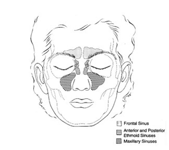
| Drawing | Sinuses |
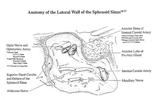
| Drawing | Sphenoid Sinus |
| Location | |
| Image5 | Identify the Sinuses you see |
| Image6 | Identify the Sinuses you see |
| Image7 | Identify the Sinuses you see |
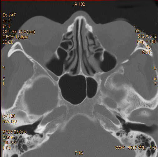
| Image 1 | Identify Maxillary Sinus |
| Image 2 | Identify Maxillary and Mastoid sinus |
| Image 3 | Identify Maxillary, Mastoid, Sinuses sphenoid sinuses |
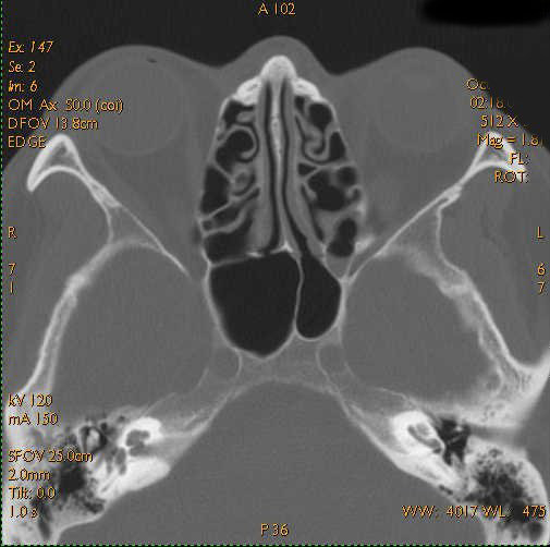
| Image 6 | Identify Ethmoid Sinus |
| Image 7 | Identify Ethmoid sinus |
| Image 8 | Identify Ethmoid sinus |
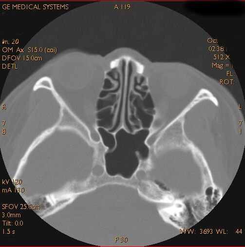
| Image 11 | Identify Frontal and Maxillary sinus |
| Image 13 | Locate Sphenoid sinus |
| Image 14 | Identify Sphenoid Sinus |
| Walls of Sinus | |
| Drainage site | |
| Image 4 | Locate the drainage site of Maxillary Sinus |
| Image 9 | Drainage site for Ethmoid sinus LABELED IMAGE NEEDED |
| Image 12 | Drainage site for Frontal sinus LABELED IMAGE NEEDED |
| Image | Drainage site for Sphenoid sinus |
| Relationships | |
| Image 5 | Appreciate the relationship of Maxillary sinus to Orbit and Teeth |
| Image 10 | Note the relationship of Ethmoid sinus to Sella Tursica |
| . | Appreciate the relationship of Frontal sinus to |
| Image 15 | Appreciate the relationship of Sphenoid sinus to Pituitary fossa. LABELED IMAGE NEEDED |
| Image 16 | Nasal turbinates, Maxillary, Ethmoid sinus LABELED IMAGE NEEDED |
| Image 17 | Note the relationship of Turbinates to ostea where sinuses drain. |
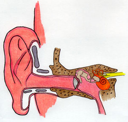
| Illustration | Auditory system |
| External auditory canal | |
| Image 2 | Identify external auditory meatus |
| Tympanic Membrane | |
| Image 9 | Locate Tympanic membrane |
| Image 10 | Locate Tympanic membrane |
| Internal auditory canal | |
| Image 3 | Identify internal auditory canals |
| Image 6 | Identify Internal auditory canal /meatus |
| Middle ear | |
| Image 1 | Identify semicircular canals |
| Image 5 | Identify malleus and cochlea |
| . | Identify Stapes |
| . | Identify Incus |
| Auditory Nerve | |
| . | Identify auditory nerve |
| Relationships | |
| Image 4 | Identify the attic (antrum) |
| Image 7 | Appreciate the relationship of middle ear to Mastoid air cells |
| Image 8 | Locate Eustachian tubes |
| Image 11 | Locate Right Eustachian tube and External auditory canal |
| Image 12 | Locate Eustachian tube |
| Anatomy | Nasal turbinates and drainage site of Maxillary sinus |
| Nasal Bones | |
| Image 1 | Identify the Nasal bones |
| Image 2 | Identify Nasal bone and bony septum |
| Nasal Septum | |
| Image 5 | Nasal septum |
| Image 4 | Identify Nasal septum |
| Nasal Fossa | |
| Image 3 | Identify the Nasal fossa and bones |
| Image 10 | Appreciate the direction of Nasal passage |
| Nasal Turbinates | |
| Image 6 | Identify Nasal Turbinates |
| Nasal Concha | |
| Drainage Sites of sinuses | |
| Image 7 | Locate the drainage site of Maxillary Sinus |
| . | Identify drainage site of Frontal sinus |
| . | Identify drainage site of Ethmoid sinus |
| Relationships | |
| Image 8 | Note the relationship of Nose to Orbit and Sinuses |
| Image 9 | Identify Nasal concha, nasal septum and see the relationship to nasopharynx and maxillary sinus |
|
Eye |
|
| Image 1 | Identify orbital fossa |
| Identify walls of orbit | |
| Image 2 | Identify Globe |
| Image 3 | Identify Lens |
| Image 4 | Identify Lateral and Medial Rectii |
| Image 5 | Identify Optic nerve |
| Image 9 | Eye, Optic nerve, optic foramen, medial and lateral recti, lens |
| Relationship | |
| . | Identify the sinuses around orbit |
| Image 6 | Appreciate the relationship of Orbit to Ethmoid sinus |
| Image 7 | Appreciate the relationship of Maxillary sinus to Orbit |
| Image 8 | Note the relationship of Orbit to Sinuses and Nose |
|
Salivary glands |
|
| Image 1 | Identify Parotid gland |
| Image 2 | Locate Parotid gland |
| . | Identify Lachrymal glands |
| Image 3 | Identify Submandibular gland |
| Image 4 | Tongue, Submandibular gland |
| Nasopharynx | |
| Image 2 | Identify Nasopharynx |
| Image 3 | Identify Nasopharynx |
| Image 4 | Identify Nasopharynx, Turbinate and Maxillary sinus |
| Image 5 | Locate Nasopharynx, Hard palate and Eustachian tube |
| Image 6 | Identify Eustachian tubes connecting to Nasopharynx. |
| Image 14 | Appreciate the structures around Nasopharynx |
| Oropharynx | |
| Image 1 | Identify Nasopharynx, Pharynx, Larynx and Trachea |
| Image 7 | Identify Pharynx |
| Image 8 | Identify Maxilla and Pharynx |
| Image 9 | Identify Uvula and Epiglottis |
| Image 11 | Identify Pharynx, valleculae, epiglottis |
| Image 12 | Pharynx, valleculae, epiglottis, hyoid, mandible |
| Image 13 | Identify Para pharyngeal space |
| Image 18 | Locate Epiglottis |
| Image 19 | Locate Vallecula and Epiglottis |
| Image 16 | Locate Uvula and Pharynx |
| Image 17 | Identify Tongue and Pharynx |
| Image 20 | Identify Aryepiglottic fold |
| Image 21 | Locate Hyoid, Vallecula and Pyriform sinus |
| Image 22 | Locate Pharynx |
| Image 23 | Locate Vallecula and Pyriform sinus |
| Image 24 | Locate Vallecula and Pyriform sinus |
| Image 14 | CT:Identify Pyriform sinus and Hyoid |
| Image 15 | CT:Identify Pyriform sinus |
| Larynx / Trachea | |
| Image 10 | Identify Tongue, Nasal concha and Thyroid cartilage |
| Image 1 | Pharynx, Larynx Trachea |
| Image 2 | Identify Glottis, Thyroid cartilage and Arytenoids |
| Image 3 | Locate Thyroid cartilage and Hyoid |
| . | Identify Vocal cords |
| Thyroid cartilage | |
| Image 4 | Identify Thyroid, Sternomastoid and Steno hyoid muscles |
| Image 5 | Identify Thyroid cartilage and Arytenoids |
| Cricoid Cartilage | |
| Image 6 | Identify Cricoid cartilage |
| Image 7 | Identify Cricoid cartilage |
| Image 8 | Identify Cricoid cartilage |
| Image 9 | Identify Thyroid, Larynx and Cricoid |
| Thyroid Gland | |
| Image 10 | CT: Locate Thyroid gland |
| Image 11 | CT:Identify Trachea and Thyroid |
| Image 124 | CT:Identify the isthmus of Thyroid gland |
| Image 13 | CT:Locate Thyroid gland |
| Image | US: Identify parts of Thyroid gland |
| Image | US: Identify the vessel lateral to Thyroid gland |
| Trachea | |
| Image 16 | Identify Thyroid cartilage, Trachea and Esophagus |
| Image 17 | Epiglottis, Thyroid, Cricoid |
| Image 1 | Identify Mandible |
| Image 2 | Identify medial and lateral plate pterygoids |
| Image 3 | Identify clivus |
| Image 4 | Identify the structures |
| Image 5 | Identify the structures |
| Image 6 | Identify Mastoids |
| Image 7 | Identify Odontoid and atlanto axial joint |
| Image 8 | Inter maxillary suture |
| Image 9 | Identify hyoid and mandible |
| Image 10 | Identify the bony structures |
| Image 11 | Identify Sella Tursica |
| Image 12 | Identify Zygoma |
| Image 13 | Identify Mandible and Pterygoids |
| Image 14 | Identify hard palate, mandible, styloid |
| Image 24 | Identify Anterior Clinoid |
| . | Identify Petrous bone |
| Image4 | |
| Image 25 | Locate Crista Galli |
|
Muscles |
|
| . | Identify Scalene muscles |
| Image 1 | Identify the Masseter muscles |
| Image 2 | Identify Sterno cleido mastoid muscles |
| Image 3 | Identify medial and lateral recti |
| Image 4 | Parotid, Para pharyngeal muscle, longus coli, medial lateral pterygoid muscle, middle concha |
| Spare Images | |
| Image 9 | Identify Ethmoid sinus |
| Image 11 | Locate ethmoid sinus |
| Image 14 | Identify Frontal sinus |
| Image 15 | Identify Frontal Sinus |
| Image 19 | Locate Sphenoid sinus |
| Image 21 | Identify Ethmoid and Maxillary Sinus |
| cte_sinus_axial2 | . |
| cte_sinus_axial3 | . |
| cte_sinus_axial4 | . |
| Image 6 | Identify malleus and cochlea |
| Image 8 | Identify Mastoid air cells |
| Image 6 | Identify drainage site of Maxillary sinus |
| Image 1 | Identify nasal septum |
| Image 2 | Identify Nasal turbinates |
| Image 10 | Identify Thyroid cartilage |