|
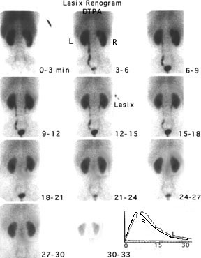
After the venous injection of radioisotope, images are collected over three minute intervals. DTPA is one of four types of radionuclide agents used in imaging of the kidney. It is filtered through the glomerulus so it is useful in detecting perfusion of the kidney and glomerular filtration. It does not demonstrate function of the renal tubules. The images are read from left to right and from top to bottom. Note that, unlike x-rays, nuclear renograms show the left kidney on the left side (as you face the scan). The data collected by the camera is analyzed by a computer and plotted on a time graph. Counts (how much isotope is in the kidneys) is shown on the Y axis and time from injection is shown on the X axis. The response of each kidney is plotted separately. Notice that both kidneys take up the isotope rapidly (the curves are steeply rising between 1 and 3 minutes). The concentration of the isotope peaks at 4-6 minutes and then starts to fall. Normally at least half of the isotope is excreted and drained from the kidneys within 20 minutes. When obstruction is suspected, a diuretic is given 15 minutes after injection of the isotope to increase urine production. This will wash the isotope out of an enlarged kidney pelvis as long as there is no obstruction to urine flow into the bladder. See a scan showing obstruction. |
|