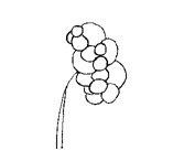
This drawing shows the atretic ureter (it is small) without a lumen attached to a collection of cysts (grossly dilated nephrons).
You are asked to see a full-term newborn female. During the pregnancy, a pre-natal ultrasound showed a cystic mass in the retroperitoneum on the left side. See the ultrasound of the mass.
The baby appears to be healthy. Her abdomen is somewhat full and there is a palpable mass in the left upper quadrant. Otherwise, the child is normal. An ultrasound obtained after the baby is born shows multiple cysts of varying sizes in the left retroperitoneum. There is no visible renal parenchyma. How can you tell whether this is a multi-cystic kidney or an obstructed kidney with hyrdonephrosis?
|
|
|
|
This drawing shows the atretic ureter (it is small) without a lumen attached to a collection of cysts (grossly dilated nephrons).
|
|
Return to Normal Renal Development.
Return to G/U Development Home Page.
©David A. Hatch, M.D., 1996