| SPACE OF DISSE WITH ITO CELLS, PITT CELLS AND RETICULUM |
|
|
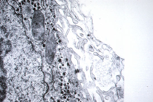
Space of Disse |
Fig 18 - A space that can be visualized only by electron microscopy between endothelial sinusoidal wall and hepatocyte.Numerous villi of hepatocytes float in this space obviously to increase their absorptive capacity. Only plasma filtrated by the endothelium flows in it. No blood cells. It contains different types of collagen,mainly type I and IV with proteoglycans an fibronectin.
|
|
|
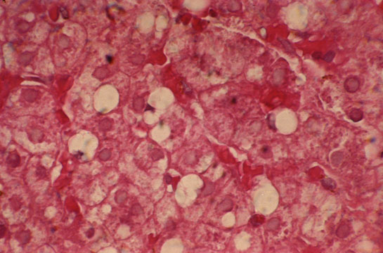
Lipocytes |
Fig 19 - ITO CELLS (Lipocytes) They are perisinusoidal cells more frequent in zone 3. They have fat vacuoles and indented nuclei.Positive for desmin under immuno stain, especially on frozen sections. They store vitamin A and produce perisinusoidal reticulum. They become hyperplastic in hypervitaminosis A. Pitt cells Are hepatic Natural Killer cell present in rat liver sinusoids. They have a higher level of activation and different morphology than NK cells of the blood.They are large ,granular lymphocytes in contact with endothelial and Kupffer cells and can be identified with electron microscopy. Their role is probably a defence against viruses and tumors. |
|
|
PITT CELLS:Are hepatic Natural Killer cells present in rat liver sinusoids.When compared with NK cells of the
blood,thy have a higher level of activation and a different morphology.They are large ,granular lymphocytes
in contact with Kupffer cells or endothelial cells.They can be identified by electron microscopy.Their role is probably
a defence against viruses and tumors. |
|
|
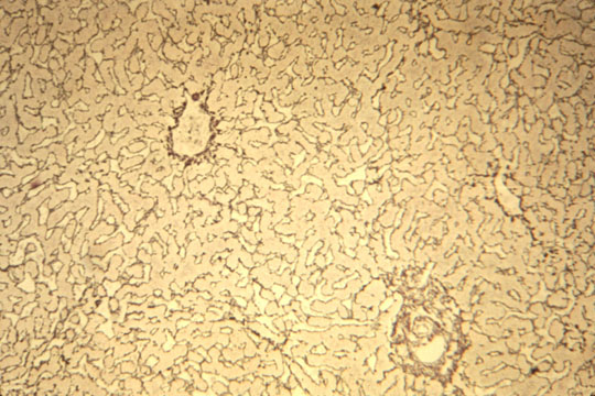
Reticulum |
Fig 20 - RETICULUM:Stacks of collagen fibers in the Disse space.They stain black with silver impregnation.Is apparently produced by Ito cells.It forms the supporting framework of the hepatocytes.Its preservation in morbid conditions permits complete regeneration ad integrum while its destruction will produce scars,fibrosis and cirrhosis.
|