LIVER CIRRHOSIS


PATHOLOGY

This peculiar transformation of the liver was identified by the first anatomic pathologist, Gianbattista Morgagni in his 500 autopsies published in 1761 but the name of "cirrhosis" (greek=orange color) was given by Laennec in 1826 because of the yellowish-tan color of the cirrhotic liver. Only in 1930, one hundred years later, however, the first theory as to the pathogenesis of this disorder was advanced by Roessle: parenchymal degeneration, regeneration and scarring which is now understood according to the following sequence:

INJURY
DEGENERATION
FIBROSIS
FORMATION OF FIBRO-VASCULAR MEMBRANES
PARENCHYMAL DISSECTION INTO NODULES
REARRANGEMENT OF BLOOD CIRCULATION
CIRRHOSIS
which is considered by most experts as a self-perpetuating irreversible process.

Regenerative nodules may form in the fibrous septa but they are not necessary for the histological diagnosis of cirrhosis: as nodules alone without fibrosis do not constitute cirrhosis. In this disease, the nodularity of the liver is mostly the result of fibrosis dissecting the parenchyma in small uniform acinar or subacinar nodules in micronodular types and in lobular and plurilobular large non-uniform nodules in macronodular forms. Regenerative nodules develop in the midst of scars but are a late phenomenon. They are important, however, for the advancement and neoplastic transformation of the cirrhosis.


Classification of cirrhosis
According to World Health Organization
(Anthony P.P. et al. J.Clin.Pathol. 31:395,1978)

MORPHOLOGIC: Macronodular   Micronodular   Mixed
HISTOLOGIC: Portal, Post-necrotic, Post Hepatitic, Biliary, Congestive
ETIOLOGIC AGENTS: Genetic, Toxic, Infectious, Biliary, Vascular, Cryptogenic

MORPHOLOGY OF CIRRHOSIS

GROSS INSPECTION.
Grossly, with the naked eye, a cirrhotic liver appears nodular, "hub-nailed", on the external surface and nodular on the cut surface. Variation in size, color, shape and consistency is relevant and may help in the identification of the etiology. The liver is usually indurated shrunken and yellowish-tan but it may be enlarged and yellow as in alcoholic fatty cirrhosis, rusty as in hemochromatosis or large and green as in biliary obstruction. It is usually the privilege of the surgeon to inspect the liver in vivo, therefore he must acquaint himself with the gross changes of cirrhosis and develop the ability of detecting discolorations of possible neoplastic nodules in order to obtain adequate samples for histological examination.

 
Micronodular cirrhosis

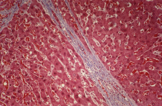
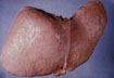
Fig.77 - MICRONDULAR CIRRHOSIS: Small rather uniform 2m nodules seperated by thin fibroussepta usually due to a chemicalagent as alcohol which diffuseuniformly throught the liver.

Macronodular cirrhosis

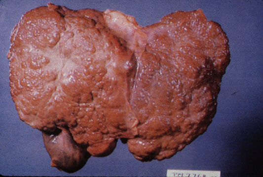
Fig.78 - MACRONODULAR CIRRHOSIS: Larger nodules separated by wider scars and irregularly distributed throughout the liver usually due to an infectious agent such as viral hepatitis which does not diffuse uniformly throughout the liver.

MICROSCOPIC CHANGES.
Presence of nodules and fibrous septa with effacement of the lobular architecture. The nodules are of two types: Dissection type and Hyperplastic Regenerative type.

Dissection nodules

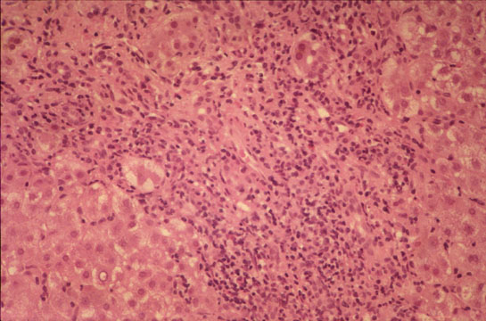
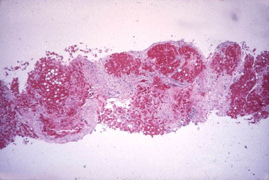
Fig.79 - DISSECTION NODULES:

  • contain remnants of portal tracts an central veins.
  • are separated by wide scars but contain thin fibrous septa.
  • contain dilated sinusoids especially at their periphery looking like multiple central veins obviously produced by the inflow of arterial blood coming from the surrounding wide scars.
  • the portal tracts within large nodules may be hypoplastic containing portal venule and arteriole but no bile ducts giving the impression of a disappearing bile duct disorder.
  • within wide scars regenerative nodules may develop.

Hypoplastic portal field

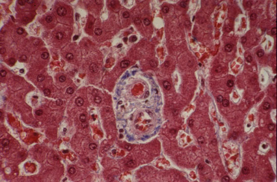
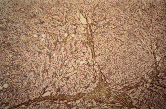
Fig.79a - HYPOPLASTIC PORTAL FIELD: In a dissecting nodule. Notice presence of portal vein, portal artery but no bile duct.This case was interpreted as "vanishing duct syndrome".

Regenerative nodules

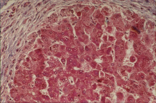
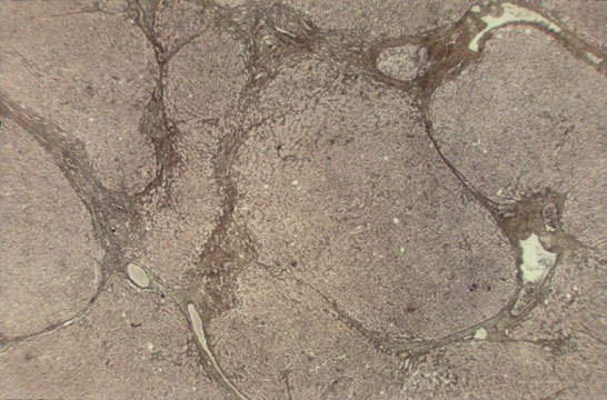
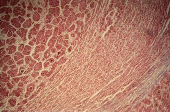
Fig.80 - REGENERATIVE NODULES:

  • these occur in micro and macro nodular cirrhosis.
  • they arise in the midst of scars favored by the rich arterial blood of scar tissue.
  • they are round nodules with a fibrous pseudo capsule with bile ductules due to obstruction of bile flow.
  • they have embryonal type of cell plates, two cells thick, "twinning of cell plates".
  • nuclei are aligned at the sinusoidal pole of the plates.
  • they often show focal cholestasis.
  • they may undergo dysplastic and malignant changes.
  • they compress the vessels of the capsule contributing to the perpetuation of the cirrhosis.


THE FIBROUS SEPTA

Are, with nodules, the other characteristic component of cirrhosis and they are visible even with the naked eye. They have been termed "fibro-vascular membranes" which provide a diversion of the blood flow through an alternative route along these fibrous septa instead of through the acinar sinusoids, thus affecting the physiology of the hepatocytes (Rappaport AM et al. virchows Archiv. A402:107-137, 1983). The fibrous septa are basically granulation tissue more or less active according to the degree of edema, capillarization, inflammatory cell infiltration and fibrosis. They reflect the activity of the cirrhotic process.
 
Passive septa

Fig.81 - PASSIVE SEPTA: Slender connective tissue bands containing few chronic inflammatory cells and sharp demarcation with parenchymal liver tissue.

Active septa

Fig.82 - ACTIVE SEPTA: Thick connective tissue bands containing edema, many chronic inflammatory cells and irregular demarcation with the parenchymal liver tissue.

  
EVOLUTION OF CIRRHOSIS

The evolution can be assessed on degree of fibrosis and nodule formation. The following stages can be identified with some approximation even on a needle biopsy specimen:


1-INCOMPLETE SEPTAL
(Incomplete bridging fibrosis, no nodules)

2-EARLY
(Thin bridging fibrosis with dissecting nodules)

3-MODERATELY ADVANCED
(Thick bridging fibrosis with dissecting nodules)

4-ADVANCED
(Wide septa with regenerative hyperplastic nodules)
  
Incomplete septal cirrhosis

Fig.83 - INCOMPLETE SEPTAL CIRRHOSIS: Presence of very slender septa radiating from enlarged fields toward the center of the lobule. There are distended efferent vessels around the septum. This type of cirrhosis produces only portal hypertension and no liver failure. The prognosis is very good if the portal hypertension is controlled.

Early cirrhosis

Fig.84 - EARLY CIRRHOSIS: Thin fibrous septa with dissecting nodules. No regenerative nodules. Presence of multiple efferent vessles. (Reticulim stain by silver impregnation).

Advanced cirrhosis

Fig.85 - ADVANCED CIRRHOSIS: Wide scars containing clusters of regenerative hepatocytes. Large scars may contain large portal fields recognizable with a stain for elastic fibers.


ACTIVITY OF CIRRHOSIS

Activity is assessed by extent of cell damage, inflammatory reaction within the scar tissue, piecemeal necrosis along fibrous septa, edema of the septa and changes in the parenchymal nodules such as necrosis and cholestasis. Activity indicates the progression of the cirrhotic process and is graded as:

INACTIVE
No inflammation and intact limiting plates around septa whiich are fibrotic

SLIGHT
Mild inflammation; segmental erosion of limiting plates

MODERATE
Moderate inflammation and damage of limiting plates

SEVERE
Marked inflammation, extensive damage of limiting plates, piecemeal necrosis
and parenchymal damage, i.e.: necrosis, cholestasis, dysplasia, malignant transformation.

COMPLICATIONS OF CIRRHOSIS

About one third of cirrhosis are compensated and, do not produce any clinical symptoms and are accidentally discovered during a medical examination or an operation or at autopsy. The rest are decompensated and produce complications mainly due to liver failure and portal hypertension. They are:

ASCITES

VARICES
Esophageal varices, hemorrhoids

PORTAL VEIN THROMBOSIS

DIGESTIVE HEMORRHAGES

JAUNDICE

HEPATIC ENCEPHALOPATHY

IMPAIRED COAGULATION

ANEMIA

INFECTION

INFARCTION OF NODULES

MALIGNANT CHANGE (HEPATOCELLULAR CARCINOMA) IN NODULES


Malignant transformation

Fig 86 - MALIGNANT TRANSFORMATION: of a regenerative nodule. Notice the capsule, the peripheral zone of the regenerative nodule and the inner zone of hepatocellular carcinoma.


LIVER BIOPSY IN CIRRHOSIS

It may assess:

PRESENCE

TYPE

ACTIVITY

EVOLUTION

CAUSE

MALIGNANT COMPLICATION

Underdiagnosis is more frequent than overdiagnosis due to the frequent absence of regenerative nodules. A pathology report should include the above parameters and known clinical complications. Treatment and prognosis depend on the correct assessment of these paramenters.

CONTENTS