 |
globules diastase PAS stain
( from College of
Amer. Pathologists)
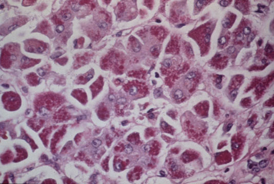
Fig 30 - ALPHA-1-ANTITRYPSIN STORAGE: The periportal hepatocytes contain very visible citoplasmic inclusions
that are diastase resistant PAS positive and can be identified with
immunoperoxidase method as alpa-1-antitrypsin (A1AT) material.
Diastase -PAS stain should be done routinely in liver biopsies.
The inclusions are almost always present in homozygous (ZZ) or
heterozygous (MZ,SZ) phenotypes of A1AT deficiency cases.
Non-alpha-1-antitrypsin similar inclusions are occasinally seen in other
conditions,such as:post mortem material and biopsy material in cirrhosis.
Immunoperoxidase reaction for A1AT will solve the problem.
Fibrinogen storage also may mimic A1AT inclusions.It can be detected
with immunoperoxidase reaction.
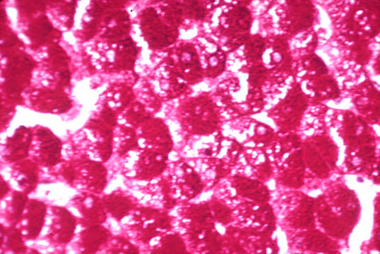 |
Fig 31 - GLYCOGEN STORAGE: On H&E stain the cytoplasm of the hepatocyte is clear and the cell membrane thick. Also the nucleus may be clear.Both cytoplasm and nucleus will be intensely positive on PAS stain,epecially in alcohol fixed specimens. These changes occur in vaious forms of hereditary glycogenosis and in acquired conditions such as diabetes,primary or induced by steroids. In diabetes there will be a large number of vacuolated noclei in the periportal area.Nuclear vacuolization ,however,may be seen without diabetes indicating that this finding is not diagnostic.It is a meaningless frequent change in autopsy material.Glycogen nuclei are very numerous and periportal in Wilson's disease.Since nucear vacuolization occurs at an early presymptomatic stage of the disease, it cannot be overlooked in a liver biopsy because it may guide to an early diagnosis and a tempestive treatment. Glycogen loss is one of the first manifestations of hepatocellular injury.
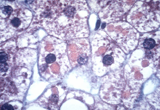 |
Fig 32 - MEGAMITOCHONDRIA: With H&E stain they show as round or elongated eosinophilic bodies with sharp borders ,containing fine granular material.They may be single or multiple and larger than the nucleus.Must be distinguished from alpha-1-antitrypsin granules and from Mallory bodies.A1AT are diastase-PAS positive.Mallory bodies have irregular margins and variable shape. Immunostains for Mallory bodies and for alpha-1-antitrypsin will be decisive.
 |
Fig 33 - GROUND GLASS HEPATOCYTES (1): Cells with a homogenous cytoplasm similar to ground glass easily spotted on H&E stain.They are seen most frequently in carrieres of HBV infection where there is marked accumulation of HBV surface antigen i the endoplasmic reticulum.. Immunostain for HBVsAg will be positive.The ground glass change,however, may occur in cases treated with various drugs which produce hyperplasia of the endoplasmic reticulum,such as barbiturates,diazepam,methotraxate, etc In this instance ,the cells are called induction cells.(2)