Ophthalmoscope should be close to your eyes. Your head and the scope should move together
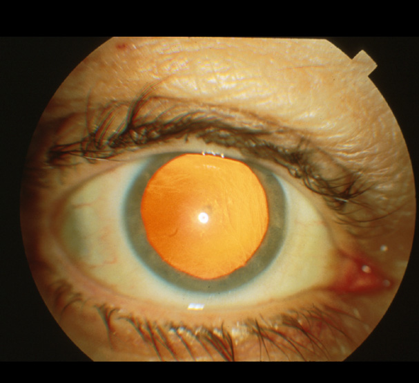
Set the lens opening at +8 to +10 diopters. With the ophthalmoscope 12-15 inches from the patient's eye, check for the red reflex and for opacities in lens or aqueous.
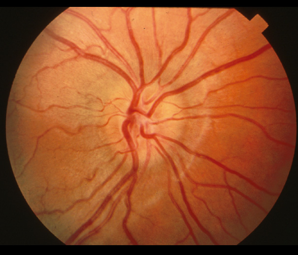
While adjusting the diopter setting, approach the patient more closely and systematically inspect the disc, noting the color, shape, margins and cup-to-disc ratio.
Inspect the vessels, noting obstruction, caliber and arterial/venous ratio.
Note the presence of arterial/venous nicking and arterial light reflex.
Check the background by inspecting for pigmentation, hemorrhages and hard or soft exudates.