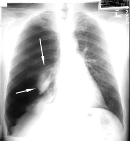
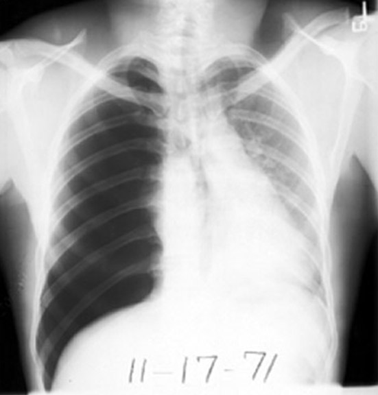
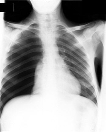
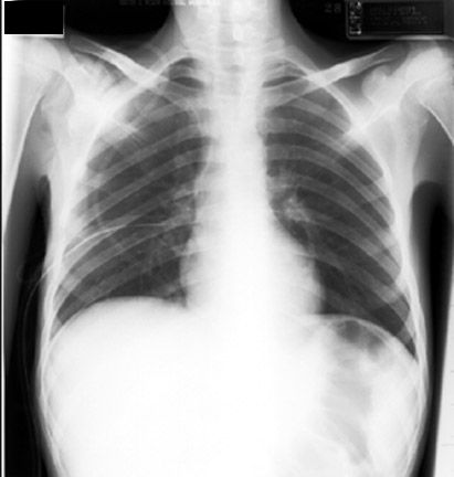
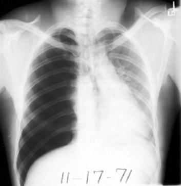
Definition: Pneumothorax is air in pleural space.
Radiological criteria:
- Air (black) in pleural space. No lung markings in pleural space.
- Recognition of atelecatatic lung (Lung
margin) The lung recoils to resting state as the negative pressure in pleura is lost (relaxation
atelectasis).
- Shift of mediastinum to opposite side. Mediastinum is held in
middle by balance between pleural pressures. when the negative pressure on the side of
Pneumothorax is lost, the mediastinum gets pulled by the normal negative pressure from the
opposite side. Progressive shift subsequently could result from a push secondary to
tension pneumothorax.
- Deep sulcus sign: The costophrenic sulcus is significantly lower than on the
contra lateral side
- Larger
hemi thorax. when the negative pressure in pleura is lost, the chest wall
reaches the TLC position. Note following
chest tube the hemi thorax returns to FRC position.
- Opposite
lung gets the entire cardiac output and the vascular markings become
prominent.
Comprehend the following:
Why does the contra lateral lung appears more dense?
What would be the radio density of the atelectatic "normal" lung
What are the true signs of tension pneumothorax?
Why is the hemi thorax larger on the side of
pneumothorax?
Does shift of mediastinum mean tension?