Endobronchial Lesion
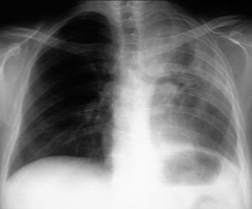 |
Left upper lobe atelectasis from endobronchial metastasis
from cancer cervix. |
- Endobronchial metastases are rare in comparison with parenchymal deposits and
account for 2% of patients who died from solid neoplasms.
- Diagnostic challenge
- They simulate primary
bronchogenic carcinoma in clinical presentation and are often difficult to distinguish,
even pathologically.
- Simultaneous occurrence of two primaries is a difficult differential
to settle on many occasions.
- The usual roentgen findings are bronchial obstruction and
obstructive atelectasis or pneumonia.
- The endobronchial lesion may have characteristic
pigment on bronchoscopy in metastatic melanoma.
- Patients may complain of persistent cough,
hemoptysis, wheezing and may have normal chest x-rays.
- Kidney, colon, breast sarcoma and
melanoma account for 67% of the cases.
- The metastases is located subepithelially
and is due to hematogenous metastases through bronchial arteries.
- It is unlikely to be
secondary to endobronchial drop metastasis as tumor cells often require fibrin thrombin to
impact. The cough and mucociliary reflex may efficiently clear aspirated cells.
- Palliative
radiation or resection becomes necessary if the patient has hemoptysis or refractory
obstructive pneumonitis.
Tracheal metastasis
When the lesion is located in the trachea, patients
will present with severe wheezing and have normal chest x-ray.