Definition
Pneumoconiosis is a group
of diseases characterized by a diffuse fibrotic reaction in the lungs induced by
inhalation of organic or inorganic particulate matter
and chemical fumes and vapors. The pathogenesis of the fibrosis is
through the release of fibrogenic chemical mediators.
Pathology
Although the end-point is fibrosis, the pattern and location varies with the type.
Coal
Workers' Pneumoconiosis: The pathology ranges from anthracosis (carbon
deposited in lung macrophages and lymphatics) to progressive massive fibrosis. The
fibrosis occurs primarily in the upper zones of the lungs.
The pathologic lesions are of 3 types
- Primary macules are <7 mm. in diameter and are composed of macrophages filled with black pigment, adjacent to a respiratory bronchiole, extending into adjacent alveoli. They are often associated with fibrosis or focal emphysema. There is only minimal impairment in lung function
- Nodular lesions are up to 2.0 cm. in diameter and are composed of interlacing
unoriented collagen bundles with dust-filled macrophages. They are not confined to the
area around respiratory bronchioles. They often encompass more proximal bronchioles and
wider zones of alveolar ducts and alveoli and other sites, particularly in subpleural and
periseptal regions
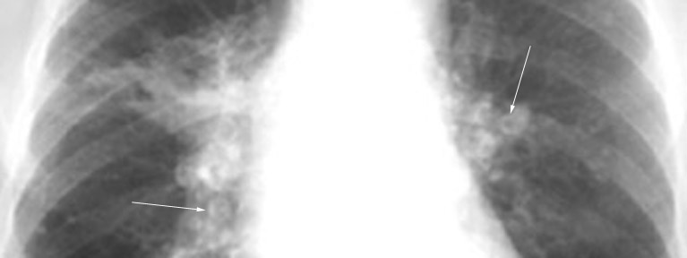
- Progressive massive fibrosis is a mass or nodule > 2.0 cm. (1 cm on x-ray)
composed of haphazardly arranged collagen with anthracotic pigment.
The center contains less pigment and is often necrotic
Silicosis:
There are 3 basic pathologic patterns of response to silica
- Fibrotic nodules are most common. They are usually <1 cm. in diameter, spherical, hard and gray to black. They are composed of concentric bundles of dense, acellular collagen. They may coalesce and have central necrosis. With central cavitation, tuberculosis should be suspected
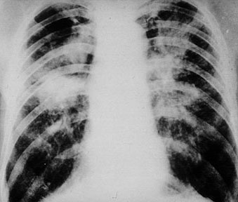
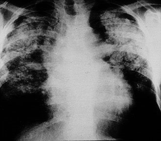
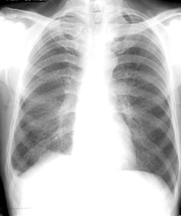
- Progressive massive fibrosis is
a form of silicosis characterized by dense agglomeration of nodules causing massive
scarring usually in the upper lobes
- Alveolar proteinosis is a pattern of lung injury caused by inhalation of large
amounts of silica. There is accumulation of proteinaceous material in the air spaces
similar to that seen in idiopathic alveolar proteinosis. However, there is more
interstitial inflammation in silicosis
Silica is ingested by macrophages. The activated macrophages release powerful
chemical mediators like interleukin-1 and tissue necrosis factor which cause fibrosis.
nodular fibrosis and larger, dense scars occur primarily in the upper zones of the lung.
Silica is present in the nodules.
Radiological features of Silicosis are:
PFT features of Silicosis
- Combined obstructive and restrictive defect.
They invariably have obstructive defect.
Occupation/Exposure for Silicosis:
- Miner
- Foundry
- Boiler scaler
- Sandblaster
Asbestosis
- The primary pathology in the lung is diffuse interstitial fibrosis which early on
predominates in the lower zones of the lungs. The histologic changes vary
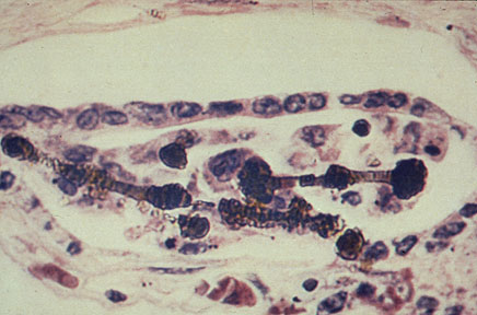
from bronchiolocentric fibrosis to honeycomb lung. The asbestos body is a unique
feature of this type of fibrosis. Asbestos bodies, which are the hallmark of exposure to
asbestos, are found within the fibrosis or alveolar spaces or occasionally in foreign-body
giant cells. An asbestos body
consists of a central core fiber of asbestos that is coated with an
iron-protein-mucopolysaccharide layer forming a golden-brown, segmented, dumb bell
appearance. Iron stains, e.g., Prussian blue, can make detection easy
- Pleural plaques (circumscribed areas of fibrosis) are the most common
manifestation of asbestosis.
- High incidence of mesothelioma and bronchogenic carcinoma.
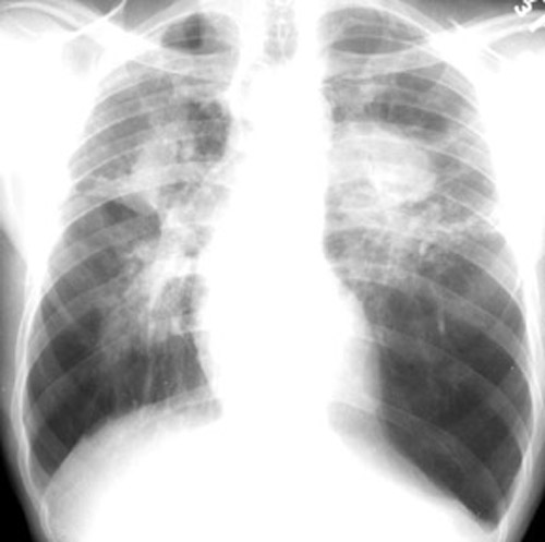
Radiological features of Asbestosis
- Pleural plaques, Calcified pleural plaques (Diaphragmatic and Parietal pleura)
- Diffuse interstitial fibrosis, Honey combing (Basal disease)
- High incidence of Lung Cancer and Mesothelioma
PFT features of Asbestosis:
Occupation/Exposure for Asbestosis:
- Shipyard
- Auto mechanics
- Insulation
Berylliosis: Beryllium induces cell-mediated immunity resulting
in non-caseating granulomas scattered throughout the lungs and hilar lymph nodes. The
granulomas "burn out," becoming fibrotic and forming fine nodular densities
throughout the parenchyma. Clinically and radiologically resembles Sarcoidosis. In
the past
Occupation/Exposure:
- Beryllium extraction
- Fluorescent light bulbs in the past
- Argon lab in the past