
|
|
Imaging |
When a patient presents with a grossly abnormal chest radiograph, has no symptoms, has a normal physical examination and no abnormal laboratory tests, sarcoidosis becomes a likely diagnosis. It should be emphasized, however, that no combination of radiograph and clinical findings are pathognomic. Sarcoidosis is a diagnosis of exclusion based on combined radiologic, clinical, and histologic data.
The radiographic abnormalities in the thorax are lymphadenopathy, parenchymal lung disease, or both. Several combinations of lymph node enlargement and lung disease have been divided into four categories or stages. These categories are of some limited clinical use since Stages I and II tend to correspond to reversible acute or subacute disease, while Stages III and IV often indicate progressive or permanent lung disease. The term stages, however, is misleading since patients do not necessarily progress from one stage to the others, and there is poor correlation between the appearance of the lung disease and functional impairment.
Stages I, II, and III are based on the chest radiograph at the time of diagnosis. Lung parenchymal disease clears completely in most patients. The development of irreversible lung disease is called Stage IV sarcoidosis.
| STAGE 0 | No Abnormality ( 5% ) |
|---|
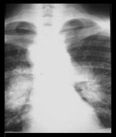
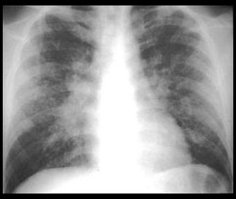
| STAGE I | STAGE II |
|---|---|
|
|
Thoracic lymphadenopathy. Normal lung parenchyma. ( 50% ) |
Hilar and mediastinal lymphadenopathy. Abnormal lung parenchyma. ( 30% ) |
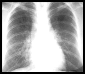
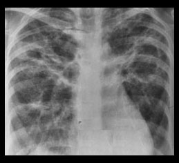
| STAGE III | STAGE IV |
|---|---|
|
|
Abnormal lung parenchyma. No lymphadenopathy. ( 15% ) |
Permanent lung fibrosis. ( 20% ) |
Reticulendothelial system
Musculoskeletal system
Gastrointestinal tract
Kidney
