Pneumoperitoneum
What are the common causes for pneumoperitoneum?
- Perforated viscus is the primary concern
- Other considerations are:
- Surgery
- Dialysis
- Toxic megacolon
- In females (intercourse, douching, insufflation)
- Pneumatosis intestinalis or coli
What imaging procedure would you order when you suspect perforated viscus?
- Upright chest x-ray
- Plain films of the abdomen upright to include the diaphragm
- CT scan would also show signs of pneumoperitoneum and the cause, such as bowel perforation.
How do you recognize pneumoperitineum in plain abdominal radiographs? What are the common imaging findings of air in peritoneum?
- An upright chest x-ray can detect as little as 1 ml of air injected into the peritoneal cavity under the diaphragm.
- Sub-diaphragmatic air
- Abdomen
- CXR upright
- Sub-diaphragmatic air
- A cross-table lateral x-ray with the patient in the left lateral position (left lateral decubitus view) can detect 5-10 ml of gas under the lateral abdominal wall.
- In a supine film, air may be seen in the sub-hepatic region.
- A supine film can show a large air collection beneath the abdominal wall that doesn't conform to any bowel loop.
- Free air can be seen under the central tendon of the diaphragm.
- Falciform ligament can be visualized in pediatric cases.
- (Rigler's sign).: Both walls of the bowel (Double wall sign) seen due to air within and outside the bowel.
- Massive air gives rise to a "Football' sign in pediatric cases.
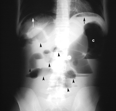
Bowel perforation / Pneumoperitoneum
Findings:
- Upright film of abdomen demonstrates air under the right hemidiaphragm (white arrow).
- Arrowheads point to multiple bowel loops with air fluid levels.
- Black arrow points to air fluid level in stomach.
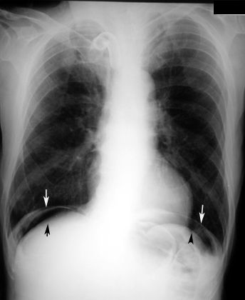
Bowel perforation / Pneumoperitoneum
Findings:
- White arrow points to diaphragm.
- Black arrow points to subdiaphragmatic air.
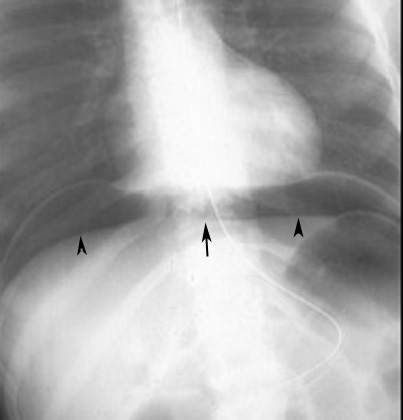
Large Pneumoperitoneum
Findings:
- Arrow points to free air central tendon
- Arrow heads pointing to free air
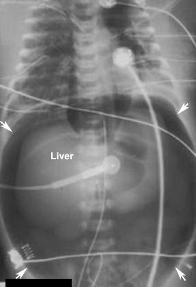
Pneumoperitoneum
Findings:
- Massive - Football sign
- Air collects under the anterior aspect of peritoneal cavity
- L: Liver
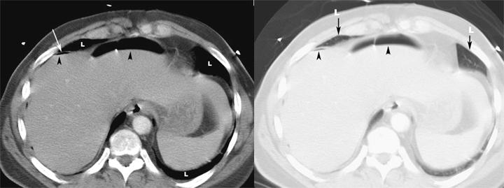
Appearance of Free Air in CT Abdomen:
Bowel perforation / Pneumoperitoneum |
|
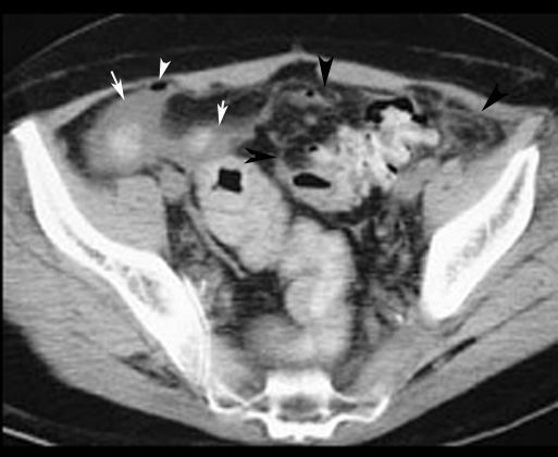
Pneumoperitoneum
- Arrowheads point to free air.
- Arrows points to collection of fluid around bowel loops.
- Black arrows point to pericolonic fascial infiltration consistent with abscess.